Already registered your school? Click here to purchase access.
View more high school resources
Step 1
Register school.
Use form on this page.
Step 2
Download and complete parent-guardian – student authorization form.
When you have received the signed authorization forms back, send notice by clicking here.*)
*We (of course) do not need to see the signed authorization form and would never ask for them.
Step 3
Purchase access.
$49/1-day access to online field trip content
Need 2 days in a row? Click here.
Need more than 2 days in a row? Contact us.
We will verify your school registration and confirm completion of parent-guardian – student authorization forms. Upon verification, we will send you login details you may use to begin the event at any date of your choosing.*
*Your login details will not expire. They will sit there ready for you until you decide to run the event. Please allow seven (7) days between notification of completion of parent-guardian – student authorization forms and receipt of login details. This means that if you plan to run the event on a Thursday, we will need to receive notice your forms are completed by the prior Monday. Please plan accordingly when distributing forms and requesting their return to you. Please also allow us one business day for other tech support responses. In other words, if you have your login and are planning to use it on Wednesday, please contact us by Tuesday rather than on Wednesday morning. Access to the field trip content is at the discretion of autopsy.online. Schools deemed unready for this content will not be granted access, and, if need be, payment refunded. If access is granted, payment is non-refundable. Access is only to the field trip content. Access to the rest of the autopsy.online website beyond the field trip content is not included with this purchase. Please also
Event description.
View a guided autopsy in the classroom. Experiencing like-there videography from the perspective of the physician performing the case. Students will feel as though they are performing the case themselves. Learn about autopsy science, medicine, surgery, and anatomy directly from patient cases.
The cost is $49 and includes a parent-guardian – student authorization form and hundreds of guiding questions to accompany the video segments. The event can be arranged anytime at your convenience. You’ll have access for one login for 24 hours and can run the event multiple times throughout the school day. (Please see below for additional tech details.)
Total video run time: 40:59. Case videos divided into short sections to allow for discussion between each section.
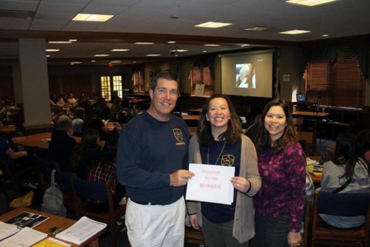
Lincoln Way East High School
NGSS-aligned
Science and Engineering Practices:
Asking questions and defining problems
Analyzing and interpreting data
Engaging in argument from evidence
Crosscutting Concepts:
Cause and effect
Systems and system models
Structure and function
Disciplinary Core Ideas:
HS-LS1-2 From Molecules to Organisms: Structures and Processes
LS3.B: Heredity; inheritance and variation of traits
What people say about our high school event
-Kelley Pataky, Science Teacher, Arlington Heights
“I really appreciate you sharing your expertise with us & giving a small look into what you do! I wasn’t expecting such a high level of humanistic concern integrated with anatomy & physiology…It was one of the best field trips I have taken my students on, certainly something they will never forget. A few months ago, we went to an anatomy lab, the students who wanted to have a more hands on experience were able to. That experience should have lessened the shock for many of the students I brought with.”
-Chad Robson, Science Teacher, Tinley Park
“…The experience was great. It was great to be able to watch a procedure such as an autopsy and then have commentary from the doctor that actually performed the procedure.”
–Nina P, student
“I also thought it was cool when he talked about the pacemaker and the steps that they had to take to make sure that they wouldn’t be shocked. There is definitely a lot of small things they have to think about when performing something so serious!”
-Jamie B., student
“[I]t was the first time I was exposed to a situation like the one we saw on screen, and it was a learning experience in and of itself.”
-Jiaqi C., student
Details
However, we will support your experience so that you and your school are ready to run the event and lead discussions.
Field Trip includes:
- Basic chest and abdomen autopsy
- 12 videos plus lung digitized histology.
- Each video with guiding discussion questions.
- 29:05 total minutes of video.
- Basic brain autopsy.
- 3 videos.
- Each video with guiding discussion questions.
- 11:54 total minutes of video.
- Parent-student authorization form.
- One login access which may be used on one device at a time only.
- Additional purchases of separate logins are required if the school plans on running the event on different devices at the same time.
- 24-hours access.
- You may preview the event the afternoon or evening before.
- How it works:
- Pick your event date.
- Set aside time the afternoon or evening the day before the event.
- Start your login access sometime after the end of your school day (the day before the event).
- Your 24-hours access will continue overnight and through the end school day on the day of the event.
- For example: If your event is on a Friday and your school day ends at 3 pm, you might start your login at 4 pm on Thursday. Preview the event. You’ll be ready for Friday. On Friday, you’ll have access till 3:59 pm. Run the event as many times as wish on the day of the event. You’ll have login access for the whole school day.
Detailed instructions are provided in your confirmation email. Please check your spam folder if you do not see your confirmation email in your inbox after subscribing.
By subscribing you understand and agree
- that access will be to the above listed content only.
- that access to the full site, database, search functions, interactive features, self-assessments, etc. is not included.
- to the Terms of Use and Privacy Policy.
Please also see Preview below.
Case 1 – chest and abdomen
Brief History: The patient was an active, elderly Caucasian woman on hypertensive medication, recently hospitalized for pneumonia. She lost 10 pounds in the hospital. She completed her antibiotic course at home and was recovering, with a residual cough. She was found dead face down on the floor after an unwitnessed cardiopulmonary arrest approximately two weeks after discharge.
Part 1
Video will appear here during field trip access.
Please contact us to learn more.
Scroll down for more questions.
1. Why is the body red?
2. How does this fit for the history?
3. What do you anticipate the subcutaneous tissue will look like under the red areas?
4. Why are there EKG leads on the body?
5. Do you think the patient received CPR?
6. What could account for the bruise in the right lower abdomen?
7. Is it likely that the bruise occurred after death?
8. How decomposed is the body?
Part 2
Video will appear here during field trip access.
Please contact us to learn more.
Scroll down for more questions.
1. What is the yellow tissue under the skin?
2. What is making that tissue red close to the skin?
3. If you were to look under the microscope at the red area under the skin, what would you see?
4. What is a partial autopsy or a limited autopsy?
5. How would the incision differ in a “chest-only” autopsy? (Draw it out on a body sketch.)
6. As the video goes along, identify these structures: ribs, costochondral junction, pectoralis major muscle, pectoralis minor muscle, sternocleidomastoid muscle.
7. What anatomic layers are being cut (from internal to external) from 2:55 to 3:10 in the video?
8. Do you see a cause of death yet?
Part 3
Video will appear here during field trip access.
Please contact us to learn more.
Scroll down for more questions.
1. What is the name of the scissors with the rounded end?
2. Why is the physician using this tool?
3. What is the ileocolic or ileocecal junction?
4. What is the taenia coli?
5. What is wider, the small intestine or the large intestine?
6. What is longer, the small intestine or the large intestine?
7. Has the patient had an appendectomy?
8. Identify the liver and the spleen.
9. What does “retroperitoneal” mean. What organs are retroperitoneal?
10. What are adhesions?
11. Where are this patient’s adhesions?
12. What specific anatomy is connected by adhesions here?
13. What causes adhesions?
14. Why did this patient have adhesions?
15. What problems can adhesions cause?
16. Did the adhesions cause this patient any problems?
17. Does the patient have her uterus?
18. Identify the bladder.
19. Add to the physician’s discussion: What are some differences to between an autopsy of a man and a woman?
20. Which organs appear decomposed?
21. What is the greater omentum? Identify it in the video.
22. Why did the physician look at the skin after assessing the pelvis? What was he looking for?
Part 4
Video will appear here during field trip access.
Please contact us to learn more.
Scroll down for more questions.
1. What organs are in the chest?
2. Why is it more important to stay close to the ribs on the left side than on the right (when removing the chest plate)?
3. What are some of the initial assessments of the chest after removing the chest plate?
4. What causes pleural fluid?
5. How much pleural fluid does this patient have on the left side?
6. Does it look infected?
7. Is it transudative or exudative?
8. Do you think the pleural fluid interfered with the patient’s breathing?
9. Why did the physician put towels over the rib edges?
10. Identify the xiphoid process.
11. Identify the diaphragm.
12. Identify the pectoralis major and pectoralis minor muscles.
Part 5
Video will appear here during field trip access.
Please contact us to learn more.
Scroll down for more questions.
1. Describe the contents of the pericardial sac. Is this normal?
2. Is there evidence for tamponade? Hypertension? Pericarditis? Myocardial rupture?
3. What vessel is entered with the forceps? What is the purpose of this part of the procedure?
4. Is any pathology identified? If so, what?
5. What is the purpose of adding clamps?
Part 6
Video will appear here during field trip access.
Please contact us to learn more.
Scroll down for more questions.
1. The first incision is above the thyroid notch. What structures are being cut through?
2. What’s the difference between the pharynx, larynx, esophagus, and trachea?
3. What’s anterior, the larynx or the esophagus?
4. Identify the hyoid bone.
5. Identify the vocal cords.
6. Why does the physician keep mentioning about not cutting through the skin of the neck?
7. What is the purpose of “tying off the carotids” and when is that performed?
8. What pathology is present in the spine? What causes this?
9. What structures (tubes) pass through the diaphragm and connect the chest organs to the abdominal organs?
10. Order these structures from anterior to posterior.
11. Order these structures from right to left.
12. What structure is being cut at 2:02?
13. What structure is being cut at 2:10?
Part 7
Video will appear here during field trip access.
Please contact us to learn more.
Scroll down for more questions.
1. What is the ligament of Treitz?
2. What organ is being removed?
3. Why is it being removed?
4. What is the name of the fat being cut?
5. Is the cut mesenteric or antimesenteric?
6. What does “blunt dissection” mean?
7. Why are strings tied around intestine before cutting between them?
Part 8
Video will appear here during field trip access.
Please contact us to learn more.
Scroll down for more questions.
1. The video begins cutting tissue lateral to the bladder. The tissue connects the bladder to the pelvic side wall. What structures are in this tissue?
2. The anterior bladder is relatively free (separated) from the anterior pelvic wall. Is that the normal configuration? Or has there already been some postmortem or dissection? Or is this a post-surgical change? (If so, what surgery?)
3. The space posterior to the bladder is relatively free. Is that the normal configuration? Or has there already been some postmortem dissection? Or is this a post-surgical change? (If so, what surgery?)
4. Identify the right lobe of the liver, the round ligament, and the left lobe the liver.
5. Is the gallbladder present?
6. Can you identify the central tendon of the diaphragm?
7. Identify any iliac vessels. Are they arteries or veins?
8. Can you definitively number any thoracic vertebrae (T1, T2, T3, etc.). What landmark did you use?
9. From about 1:20 to 1:40, you can see a thin, long strand on either immediate side of the thoracic vertebral column and spanning the entire thorax. What is it?
10. Is the phrenic nerve visible in the video?
11. What are indications for checking urine for postmortem toxicology?
12. What pre-analytic variables might affect urine toxicology results?
Part 9
Video will appear here during field trip access.
Please contact us to learn more.
Scroll down for more questions.
1. Identify the right upper, middle, and lower lobes; and the left upper and lower lobes.
2. Identify the oblique fissure. Identify the bilateral horizontal fissures.
3. What is anthracosis? Does the patient have it?
4. What structures comprise the lung hila?
5. Once the hilar structures are visible on the right, identify the bronchus, pulmonary artery, and pulmonary veins?
6. Once the hilar structures are visible on the left, identify the bronchus, pulmonary artery, and pulmonary veins?
7. Did you identify the structure in 2. and 3. based on relative location or appearance?
8. Draw out the right hilar structure, correctly positioned (which is anterior, posterior, medial later). Repeat for the left.
9. Which is thicker and firmer, the pulmonary artery or vein?
10. Which vessel is the pathologist checking for pulmonary embolism?
11. What is the difference between a central and peripheral pulmonary embolism?
12. Is it possible to have microscopic pulmonary embolism?
13. Did this patient have a pulmonary embolism?
14. Was the patient a smoker? What anatomy evidence is there for your answer?
15. There is a lymph node identified. Does it appear normal or diseased? How can you tell?
16. Where are thoracic lymph nodes typically found (named areas)?
17. What are the lung weights? Are the lung weights normal?
18. Which lung is heavier? Is that normal?
16. What would be the clinical significance of a pulmonary embolism obstructing the right pulmonary artery vs. a pulmonary embolism obstructing a single sublobar pulmonary artery vs. a single microscopic pulmonary embolism?
19. What is acute chest syndrome in sickle cell disease?
8. How decomposed is the body?
Part 10
Video will appear here during field trip access.
Please contact us to learn more.
Scroll down for more questions.
1. What kind of pathology can be identified on the cut surface of the lungs?
2. What are examples of focal pathology in the lungs (diseases that affect one area)?
3. What are examples of diffuse pathology in the lungs (diseases that affect the entire lung)?
4. Are there any conditions that can be both focal and diffuse?
5. What main pathophysiologic process does this patient seem to have?
6. Is there pneumonia so far?
7. What would the cut surface of a lung with emphysema look like?
Part 11
Video will appear here during field trip access.
Please contact us to learn more.
Scroll down for more questions.
1. Identify the heart, aorta and larynx.
2. Identify the left and right sides of the heart.
3. Why is one side thicker than the other?
4. Identify the esophagus.
5. Where is the esophagus in relation to the airway.
6. Identify the trachea, tracheal rings, and posterior tracheal membrane.
7. Is the trachea opened from the anterior or posterior side?
8. How decomposed is the specimen?
9. Identify the tracheal bifurcation.
10. Identify the esophageal mucosa.
Part 12
Video will appear here during field trip access.
Please contact us to learn more.
Scroll down for more questions.
1. Identify the aorta.
2. How much plaque is on the aorta?
3. Identify the main branches of the aortic arch.
4. Identify posterior intervertebral artery ostia in the aorta.
5. Identify the pulmonary artery, and the aorta.
6. What artery runs immediately posterior to the origin of the pulmonary artery.
7. Identify the left anterior descending coronary artery.
8. Is there any plaque in it? How much? Is it enough to present a cause of death?
Part 13
Video will appear here during field trip access.
Please contact us to learn more.
Scroll down for more questions.
1. Review lung histology.
2. Identify alveoli (alveolar walls and spaces).
3. How many alveoli have “pink stuff” inside them?
4. How many alveoli are “clear” inside?
5. What do you think the “pink stuff” is inside the alveoli?
6. What is normally inside the alveolar spaces in healthy people?
7. Should alveoli in healthy people be clear or have something inside them?
8. Review the circulatory system — specifically how blood circulates between the heart and lungs.
9. What is pulmonary edema?
10. How does pulmonary edema happen?
11. Why did the patient die?
Case 16 – brain
Brief History: The patient was an elderly Caucasian man, status post kidney transplant who had longstanding undiagnosed dementia.
Part 1
Video will appear here during field trip access.
Please contact us to learn more.
Scroll down for more questions.
1. What is the purpose of the brain removal in this case?
2. Why part the hair?
3. What layers of the scalp are being separated manually (with the fingers)?
4. Identify the temporalis muscle.
5. What does “reflect” mean in an autopsy? For example, what does it mean to “reflect skin” or “reflect muscle”?
6. What’s the purpose of reflecting the temporalis muscle?
7. What are the attachments of the temporalis muscle?
8. Why is there no bleeding during the scalp incision?
Part 2
Video will appear here during field trip access.
Please contact us to learn more.
Scroll down for more questions.
1. What muscle is folded down on the right side of the skull?
2. How thick is the normal skull?
3. Why add the notch in the bone above the ear?
4. Is the body face up or face down?
5. What layers of the skull are visible?
6. Are we looking directly at skull bone?
7. Can you identify any sutures? If not, why not?
Part 3
Video will appear here during field trip access.
Please contact us to learn more.
Scroll down for more questions.
1. Identify the epidural, subdural, and subarachnoid space near the start of the video.
2. What is the purpose of adding notches on either side of the skull?
3. Why is the spinal fluid bloody? Is this a sign of disease?
4. What gross findings can suggest dementia?
5. Can any type of dementia be diagnosed grossly? Which type(s)?
6. Which dementias require a microscopic diagnosis?
7. What structures are under the cerebellar tentorium?
8. What main structures attach the brain to the body? (Or which structures need to be cut to release the brain from the skull?)
9. Identify each structure:
Skull
Base of skull
Anterior cranial fossa
Middle cranial fossa
Posterior cranial fossa
Dura mater
Cerebellar tentorium
Temporal lobe
Frontal lobe
Parietal lobe
Occipital lobe
Pituitary stalk
Sella turcica
Clivus
Petrous bone
Sphenoid wing
Optic nerve