Case 19 Part 5
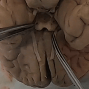
Brain — detailed external anatomy.
What’s inside? cranial nerves
. Medulla
2. Pons
3. Meningioma
4. Groove of herniation (likely from embalming, postmortem)
5. Mengioma
6. Cut surface of meningioma
7. Temporal lobe (right)
8. Frontal lobe (right)
9. Basilar artery
10. Cranial nerve III (right)
11. Olfactory nerve (right, bulb transected) cranial nerve I cranial nerve 1
12. Internal carotid artery (left)
13. Basilar artery
14. Cranial nerve VI (left) cranial nerve 6 abducens nerve
15. Right internal carotid artery
16. Optic chiasm
17. Posterior cerebral artery (right, circle of Willis)
18. Continuation of right posterior cerebral artery
19. Posterior communicating artery (right, circle of Willis)
20. Basilar artery
21. Superior cerebellar artery (left)
22. Basilar artery
23. Posterior cerebral artery (left, circle of Willis)
24. Cranial nerve II (left, optic nerve) cranial nerve 2
25. Basilar artery
26. Posterior cerebral artery (right, circle of Willis)
27. Posterior communicating artery (right, circle of Willis)
28. Junction of internal carotid and middle cerebral artery (right, circle of Willis)
29. Mamillary body (left)
30. Cranial nerve III (right) oculomotor nerve cranial nerve 3
31. Optic chiasm
32. Cranial nerve II (left, optic nerve) cranial nerve 2
33. Pyramid/medulla (left)
34. Cerebellum (right)
35. Middle cerebellar peduncle (right)
36. Pons
37. Cranial nerve IV cranial nerve 4 trochlear nerve
38. Substantia nigra (left)
39. Cerebral peduncle (right)/midbrain
40. Pons
41. Cerebral aqueduct
42. Medulla
43. Substantia nigra
44. Medulla
45. Pons
Embalmbed
Embalming
Meningioma
Brain
Cerebrum
Inferior brain
Brainstem
Circle of Willis
Mamillary body
Internal cerebral artery
Pituitary stalk
Midbrain
Pons
Medulla
Brainstem
4th cranial nerve
Cranial nerve IV
Substantia nigra
Middle cerebral peduncle
. Medulla
2. Pons
3. Meningioma
4. Groove of herniation (likely from embalming, postmortem)
5. Mengioma
6. Cut surface of meningioma
7. Temporal lobe (right)
8. Frontal lobe (right)
9. Basilar artery
10. Cranial nerve III (right)
11. Olfactory nerve (right, bulb transected) cranial nerve I cranial nerve 1
12. Internal carotid artery (left)
13. Basilar artery
14. Cranial nerve VI (left) cranial nerve 6 abducens nerve
15. Right internal carotid artery
16. Optic chiasm
17. Posterior cerebral artery (right, circle of Willis)
18. Continuation of right posterior cerebral artery
19. Posterior communicating artery (right, circle of Willis)
20. Basilar artery
21. Superior cerebellar artery (left)
22. Basilar artery
23. Posterior cerebral artery (left, circle of Willis)
24. Cranial nerve II (left, optic nerve) cranial nerve 2
25. Basilar artery
26. Posterior cerebral artery (right, circle of Willis)
27. Posterior communicating artery (right, circle of Willis)
28. Junction of internal carotid and middle cerebral artery (right, circle of Willis)
29. Mamillary body (left)
30. Cranial nerve III (right) oculomotor nerve cranial nerve 3
31. Optic chiasm
32. Cranial nerve II (left, optic nerve) cranial nerve 2
33. Pyramid/medulla (left)
34. Cerebellum (right)
35. Middle cerebellar peduncle (right)
36. Pons
37. Cranial nerve IV cranial nerve 4 trochlear nerve
38. Substantia nigra (left)
39. Cerebral peduncle (right)/midbrain
40. Pons
41. Cerebral aqueduct
42. Medulla
43. Substantia nigra
44. Medulla
45. Pons
Embalmbed
Embalming
Meningioma
Brain
Cerebrum
Inferior brain
Brainstem
Circle of Willis
Mamillary body
Internal cerebral artery
Pituitary stalk
Midbrain
Pons
Medulla
Brainstem
4th cranial nerve
Cranial nerve IV
Substantia nigra
Middle cerebral peduncle