Library
Autopsy.Online
Desktop devices
recommended.
Autopsy.Online
How would you like to search the database?
By General Case Topic 🛈
This is a brief, partial “quick-start” guide to assist with case selection. Most cases have multiple clinical and pathologic issues. Please review the history and other search options for a more detailed exploration of case content.
↓ Scroll down for more.
Basic introductory autopsy
Adult:
Fetal:
Medicine
Dialysis management, chronic
pulmonary embolism Case 27
Gastric bleeding, acuteCase 52
Gastrointestinal bleeding,
chronic Case 11
Hypertensive heart disease Case 14
Ischemic stroke Case 20
Meningioma Case 19
Mesothelioma Case 4
Multiple tumors, smoking Case 40
Renal failure Case 41
Renal failure, alcohol
use disorder Case 37
Retroperitoneal mass Case 49
Critical Care
Cardiac tamponade Case 33
Pulmonary embolism Case 12
Pulmonary embolism Case 29
Pulmonary embolism Case 42
Peritonitis Case 10
Portal hypertension Case 39
Renovascular hypertension Case 44
Shock Case 8
Surgical Complications
Coronary stent Case 34
Coronary stent Case 50
Hysterectomy Case 47
Lung mass resection Case 38
Pacemaker placement Case 46
Spinal fusion Case 6
Thrombectomy, endarterectomy Case 28
Trauma
Fall Case 36
Fall Case 43
Low-speed motor vehicle
accident Case 25
High-speed motor vehicle
accident Case 35
Forensics
By Audio 🛈
Click one or both options for cases with audio explanation or guidance.
Leave the this option blank if you don’t need site explanation:
-students doing labs
-educators making your own case interpretation
-if you are looking for cases to figure out on your own from the visual

By Visual Guide
By Case History
By Clinical Topic

More ↓
Search the History for case information.
By Case
on larger devices.)
on tablet or larger devices.)
Back to top Showing all 355 results
-

Case 1 – History
Recent pneumonia.
-
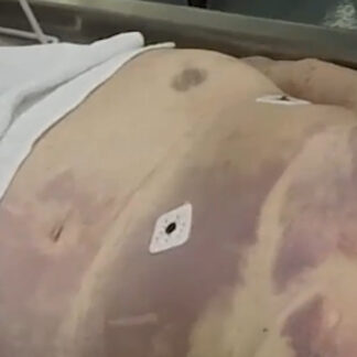
Case 1 Part 1
External exam. Anterior livor mortis. EKG leads.
-
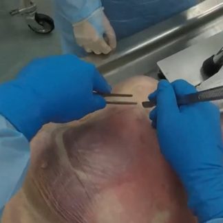
Case 1 Part 2
Y-shaped incision. Livor mortis within subcutaneous tissue.
-
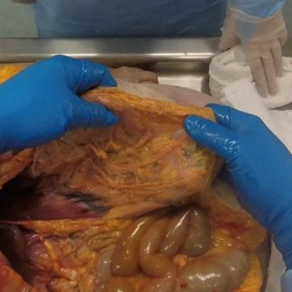
Case 1 Part 3
Abdominal survey — basic anatomy. Adhesions.
-
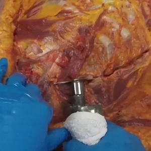
Case 1 Part 4
Removal of chest plate.
-
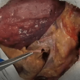
Case 1 Part 5
Assessment for pulmonary embolism.
-
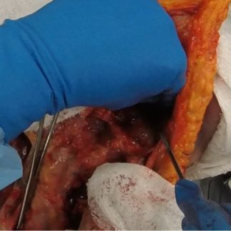
Case 1 Part 6
Neck anatomy — basic review. Removal of chest organs.
-
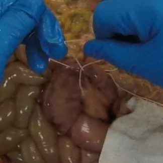
Case 1 Part 7
Removal of intestine.
-
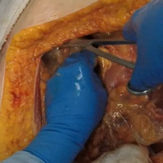
Case 1 Part 8
Removal of bladder. Status post hysterectomy. Basic survey of abdomen. Scoliosis.
-
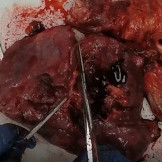
Case 1 Part 9
Heart and lung block — basic anatomy.
-
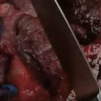
Case 1 Part 10
Lung sectioning.
-
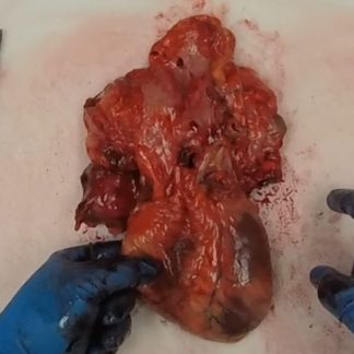
Case 1 Part 11
Airway, heart (external), esophagus — basic anatomy.
-
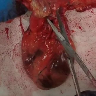
Case 1 Part 12
Lining of aorta. Left anterior descending coronary artery (plaque).
-

Case 1 Part 13
Microscopy — pulmonary edema.
-

Case 2 – History
Diabetes, stroke, feeding tube.
-
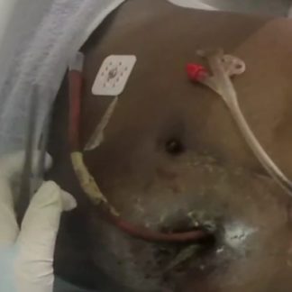
Case 2 Part 1
Gastrostomy tube. Jejunostomy tube.
-
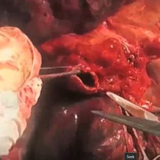
Case 2 Part 2
Aspiration.
-
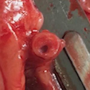
Case 2 Part 3
Severe 2-vessel coronary artery disease.
-
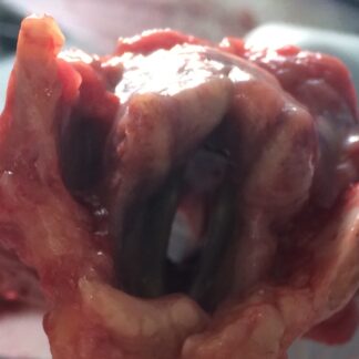
Case 2 Part 4
Laryngeal stenosis. Traumatic intubation.
-

Case 3 – History
Recent chest pain.
-
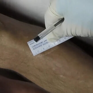
Case 3 Part 1
External exam.
-
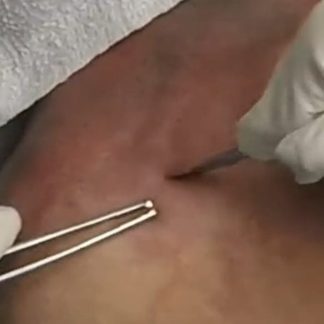
Case 3 Part 2
Y-shaped incision.
-
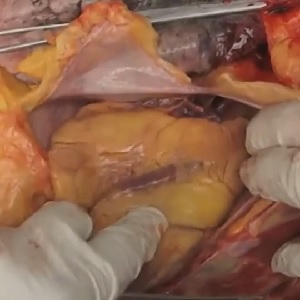
Case 3 Part 3
Initial chest assessment.
-
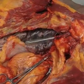
Case 3 Part 4
Continued chest assessment.
-
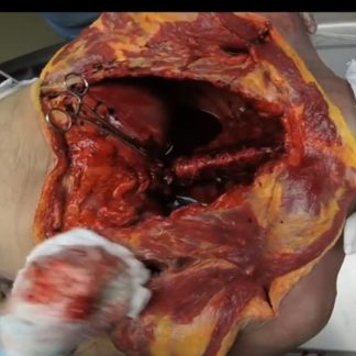
Case 3 Part 5
Organ block. Chest cavity.
-
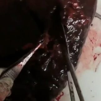
Case 3 Part 6
Pulmonary apical blebs (emphysema).
-
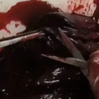
Case 3 Part 7
Lung weights. Pulmonary congestion and edema.
-
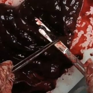
Case 3 Part 8
Lung sampling for microscopy.
-
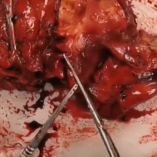
Case 3 Part 9
Airway. Aorta. Esophagus.
-
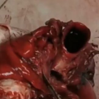
Case 3 Part 10
Assessment of coronary arteries.
-
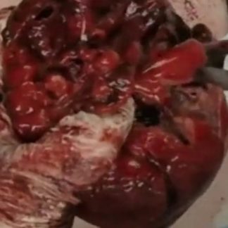
Case 3 Part 11
Left dominant coronary artery system.
-

Case 4 – History
Transdiaphragmatic tumor.
-
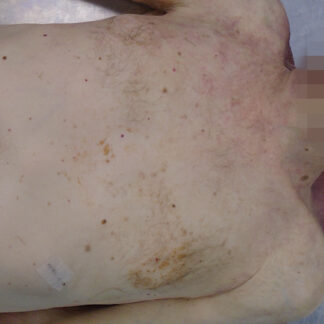
Case 4 Part 1
External exam.
-
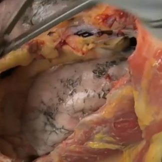
Case 4 Part 2
Transdiaphragmatic tumor. Pleural effusion.
-
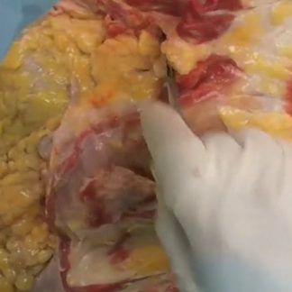
Case 4 Part 3
Tumor attached to pericardium. Pleural effusion.
-

Case 5 – History
Dementia
-
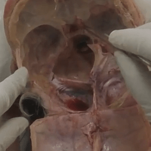
Case 5 Part 1
Base of skull anatomy.
-
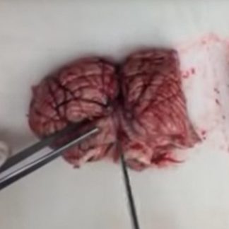
Case 5 Part 2
Cerebellar anatomy.
-
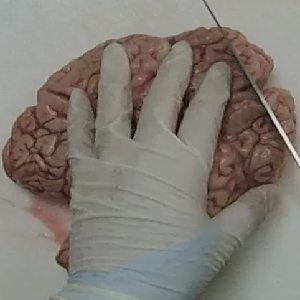
Case 5 Part 3
Cerebral anatomy.
-

Case 6 – History
Spinal surgery.
-
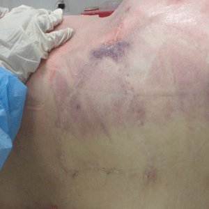
Case 6 Part 1
External exam. Multiple surgical scars.
-
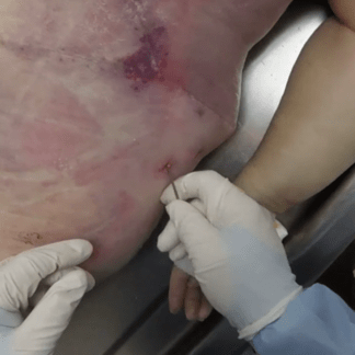
Case 6 Part 2
Spine exposure.
-
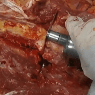
Case 6 Part 3
Bone saw.
-
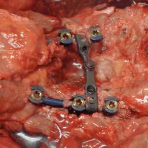
Case 6 Part 4
Spinal cage.
-
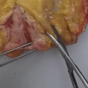
Case 6 Part 5
Right flank assessment.
-
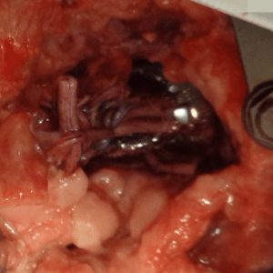
Case 6 Part 6
Spinal cord. Cauda equina.
-
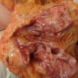
Case 6 Part 7
Abscess.
-
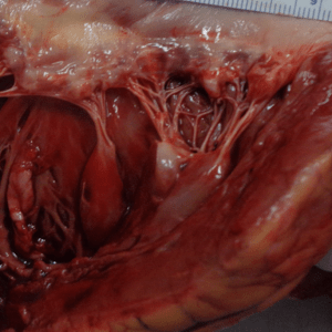
Case 6 Part 8
Heart anatomy.
-

Case 7 – History
Inferior vena cava filter.
-
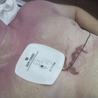
Case 7 Part 1
Rigor mortis.
-
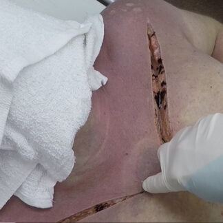
Case 7 Part 2
Y-shaped incision.
-
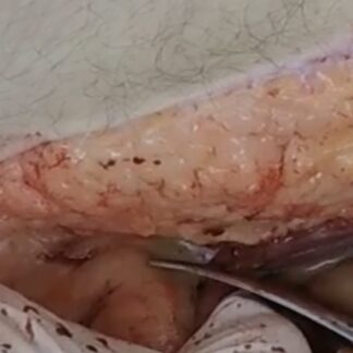
Case 7 Part 3
Initial dissection.
-
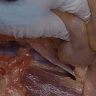
Case 7 Part 4
Ureter. Inferior vena cava.
-
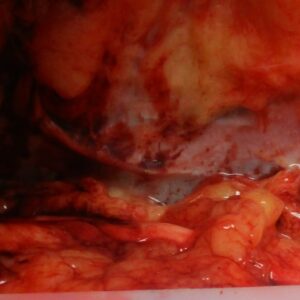
Case 7 Part 5
Inferior vena cava filter.
-
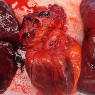
Case 7 Part 6
Heart and lung block.
-

Case 8 – History
TIPS procedure.
-
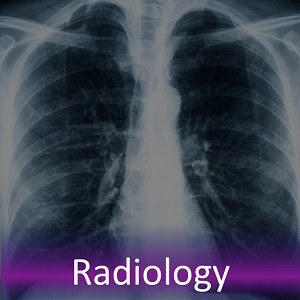
Case 8 Part 1
TIPS fluoroscopy. Postoperative CT scan.
-
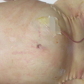
Case 8 Part 2
External exam. Peritoneal dialysis catheter. Central venous access sites. Long bone donation.
-
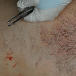
Case 8 Part 3
Bowel infarct.
-
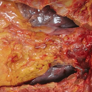
Case 8 Part 4
Pleural fluid. Pericardial fluid. Splenomegaly. Bowel infarct.
-
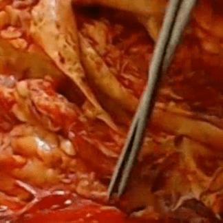
Case 8 Part 5
Aorta assessment. Dual superior mesenteric artery ostia.
-
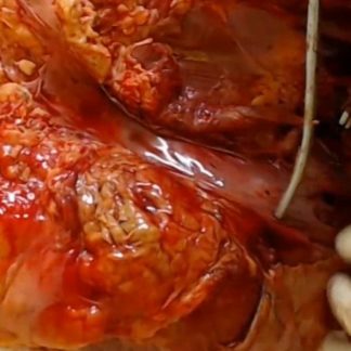
Case 8 Part 6
Inferior vena cava. TIPS device (shunt).
-

Case 9 – history
Spleen dissection.
-
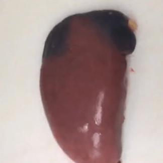
Case 9 Part 1
Spleen dissection.
-

Case 10 – History
Post-colonoscopy complication.
-
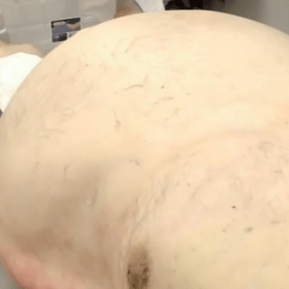
Case 10 Part 1
External exam.
-
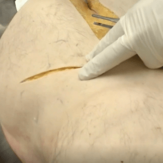
Case 10 Part 2
Ascites. Peritonitis.
-
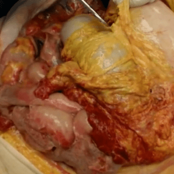
Case 10 Part 3
Peritonitis.
-
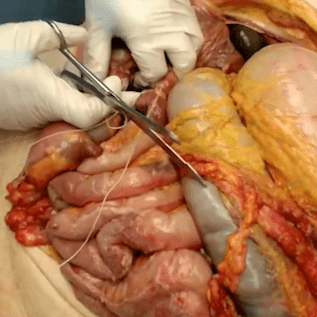
Case 10 Part 4
Peritonitis.
-
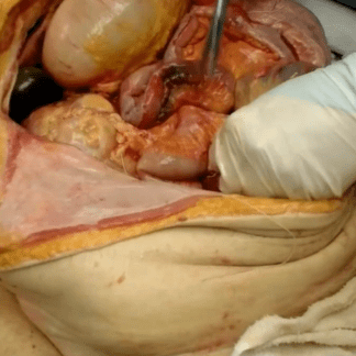
Case 10 Part 5
Peritonitis.
-
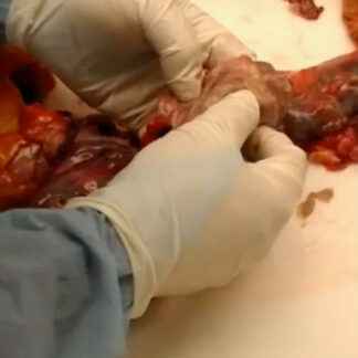
Case 10 Part 6
Assessment of large intestine.
-
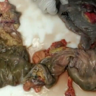
Case 10 Part 7
Biopsy site. Colonoscopy clips.
-

Case 11 – History
Gastrointestinal bleeding and sudden death.
-
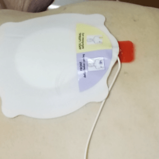
Case 11 Part 1
External exam. Left mastectomy. Obesity. Defibrillator pads. Intraosseous needle.
-
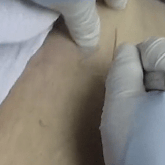
Case 11 Part 2
Y-shaped incision. Entry into abdomen. Removal of chest plate. Obesity.
-
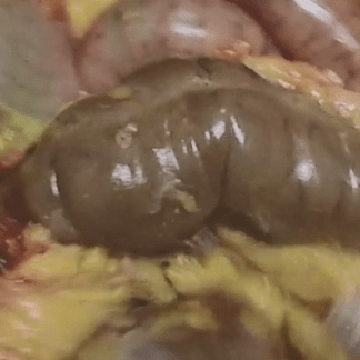
Case 11 Part 3
Abdominal dissection.
-
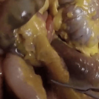
Case 11 Part 4
Removal of jejunum, ileum and large intestine.
-
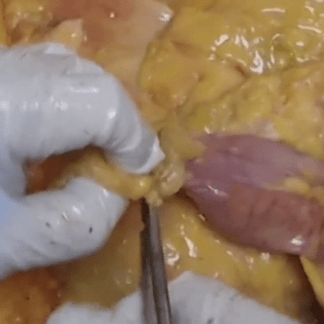
Case 11 Part 5
Removal of duodenum and stomach.
-
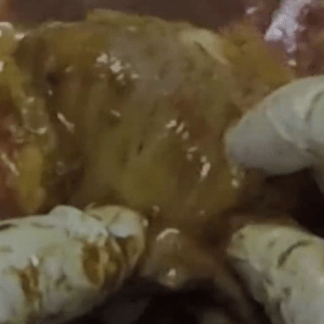
Case 11 Part 6
Diverticulosis.
-
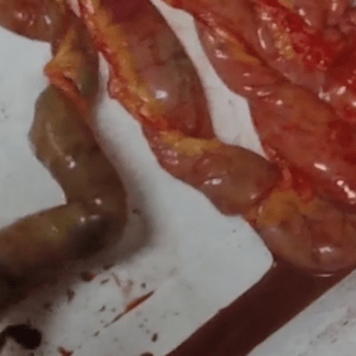
Case 11 Part 7
Acute hemorrhagic gastritis.
-
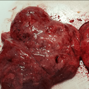
Case 11 Part 8
Pulmonary embolism.
-

Case 12 – History
Sudden death.
-
Case 12 Part 1
External exam.
-
Case 12 Part 2
Y-shaped incision.
-
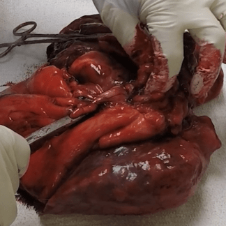
Case 12 Part 3
Heart and lung block.
-
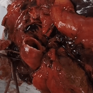
Case 12 Part 4
Acute pulmonary embolism.
-
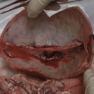
Case 12 Part 5
Removal of dura.
-

Case 13 – History
Sudden death.
-
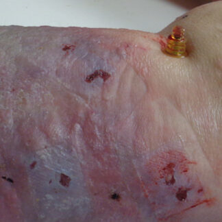
Case 13 Part 1
External exam. Intraosseous needle.
-
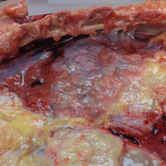
Case 13 Part 2
Adhesions.
-
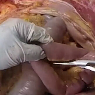
Case 13 Part 3
Assessment of abdomen.
-
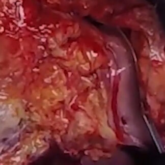
Case 13 Part 4
Chest assessment.
-
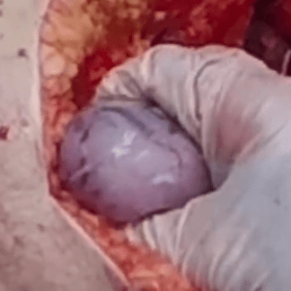
Case 13 Part 5
Testes, spermatic cord — basic anatomy.
-
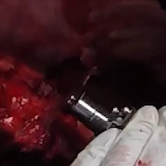
Case 13 Part 6
Lung assessment.
-
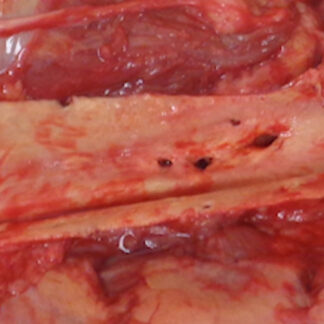
Case 13 Part 7
Aorta assessment.
-
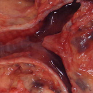
Case 13 Part 8
Airway. Posterior view. Bronchial fluid (pulmonary edema).
-
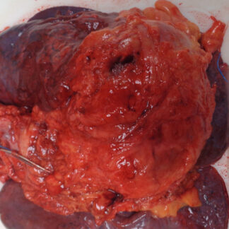
Case 13 Part 9
Coronary artery bypass graft surgery. Pacemaker-defibrillator lead insertions.
-
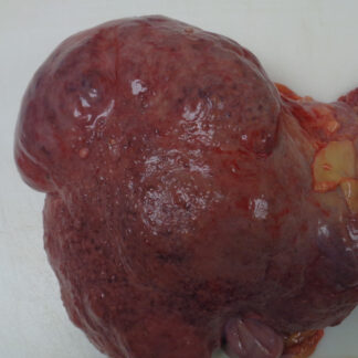
Case 13 Part 10
Cirrhosis.
-
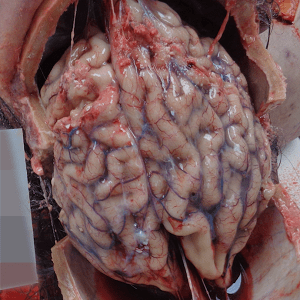
Case 13 Part 11
Brain anatomy
-

Case 14 – History
Recent chest pain.
-
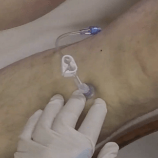
Case 14 Part 1
External exam. Intraosseous needle.
-
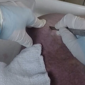
Case 14 Part 2
Y-shaped incision.
-
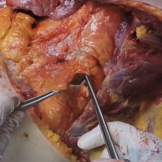
Case 14 Part 3
Chest assessment.
-
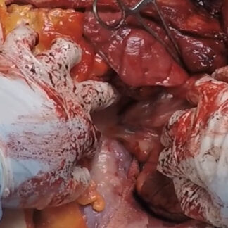
Case 14 Part 4
Chest assessment.
-
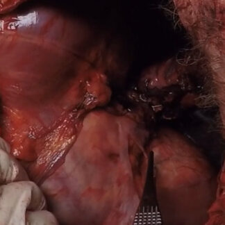
Case 14 Part 5
Diaphragm — basic anatomy.
-
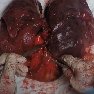
Case 14 Part 6
Heart and lung block.
-
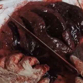
Case 14 Part 7
Lung assessment. Pulmonary edema.
-
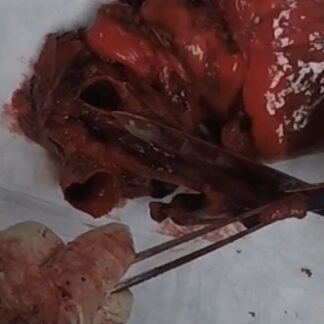
Case 14 Part 8
Lung assessment.
-
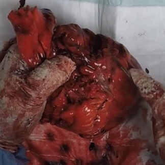
Case 14 Part 9
Coronary artery assessment.
-
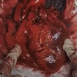
Case 14 Part 10
Left ventricular hypertrophy
-

Case 15 – History
Chronic pain and sudden death.
-
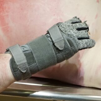
Case 15 Part 1
External exam.
-
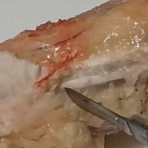
Case 15 Part 2
Distal forearm musculature — detailed anatomy.
-
Case 15 Part 3
Toxicology.
-

Case 16 – History
Dementia and kidney failure.
-
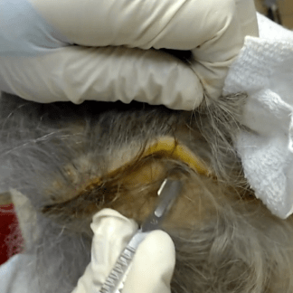
Case 16 Part 1
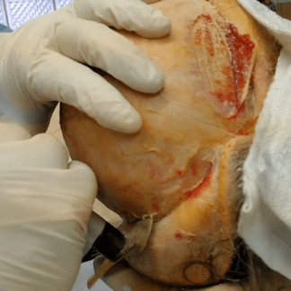
Skull exposure.
-
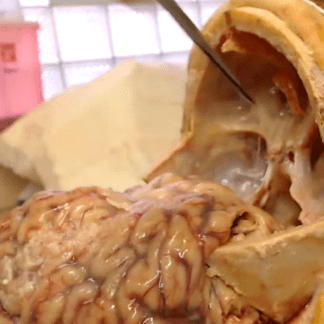
Case 16 Part 2
Brain exposure.
-
Case 16 Part 3
Brain and base of skull — basic anatomy.
-

Case 17 – History
Sudden death.
-
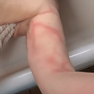
Case 17 Part 1
External exam. Early decomposition.
-
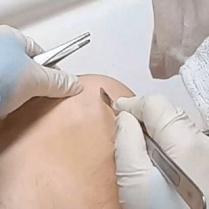
Case 17 Part 2
Y-shaped incision.
-
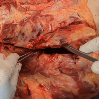
Case 17 Part 3
Rib fractures.
-
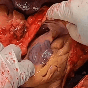
Case 17 Part 4
Chest evaluation. Pleural effusion.
-
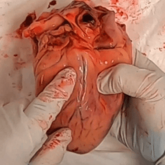
Case 17 Part 5
Heart — basic anatomy. Coronary artery blockage.
-
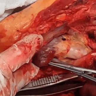
Case 17 Part 6
Endotracheal tube.
-

Case 18 – History
Decomposition. History of clipped cerebral aneurysm.
-
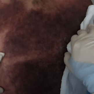
Case 18 Part 1
External exam.
-
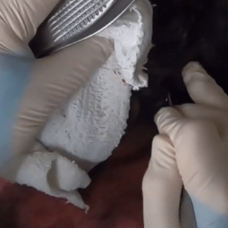
Case 18 Part 2
Skull exposure.
-
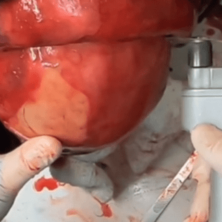
Case 18 Part 3
Brain exposure.
-

Case 19 – History
Hypertension, dizziness and sudden death.
-
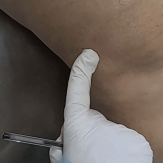
Case 19 Part 1
External exam.
-
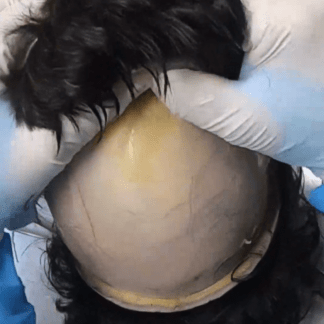
Case 19 Part 2
Skull exposure.
-
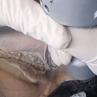
Case 19 Part 3
Brain exposure.
-
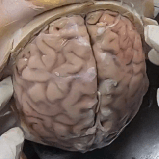
Case 19 Part 4
Brain tumor.
-
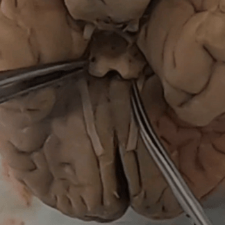
Case 19 Part 5
Brain — detailed external anatomy.
-
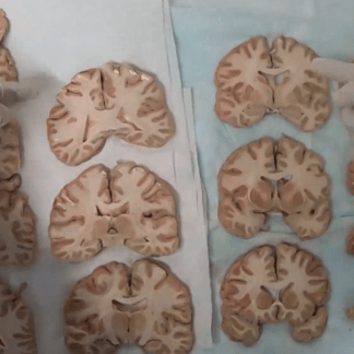
Case 19 Part 6
Brain — detailed internal anatomy.
-

Case 20 – History
Elderly man with stroke.
-
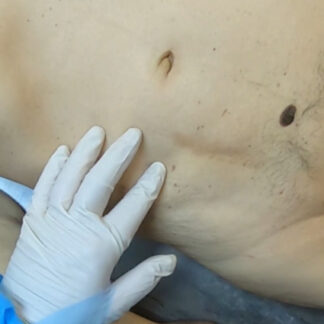
Case 20 Part 1
External exam.
-
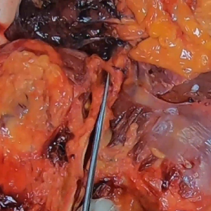
Case 20 Part 2
Abdominal aorta – main branches. Inferior vena cava. Basic anatomy.
-
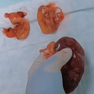
Case 20 Part 3
Gerota’s fascia. Kidney. Ureter. Adrenals. Basic anatomy.
-
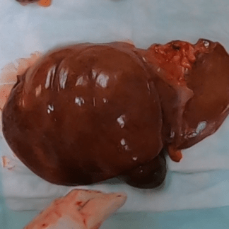
Case 20 Part 4
Liver. Gallbladder. Basic anatomy.
-
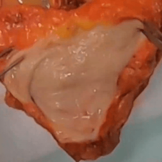
Case 20 Part 5
Ureter. Bladder. Prostate. Rectum. Basic anatomy.
-

Case 20 Part 6
Subcarinal lymph nodes.
-
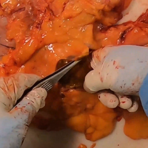
Case 20 Part 7
Duodenal mucosa. Ampulla of Vater. Basic anatomy.
-
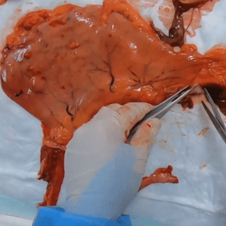
Case 20 Part 8
Gastric and esophageal mucosa. Basic anatomy.
-
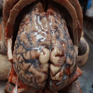
Case 20 Part 9
Old cavitary cerebral infarction.
-

Case 21 – History
History of breast cancer and alcohol use.
-
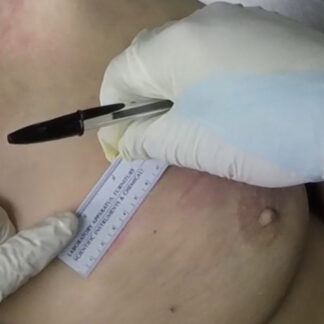
Case 21 Part 1
External exam.
-
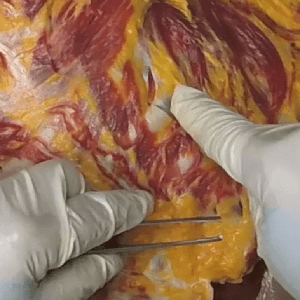
Case 21 Part 2
Removal of chest plate.
-
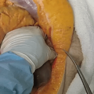
Case 21 Part 3
Abdominal survey.
-
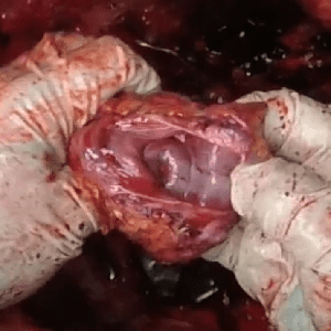
Case 21 Part 4
Uterus — basic external anatomy.
-

Case 22 – History
Middle-aged woman with psychiatric history.
-
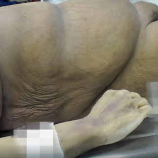
Case 22 Part 1
External exam.
-
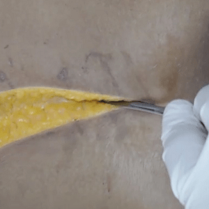
Case 22 Part 2
Y-shaped incision.
-
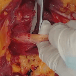
Case 22 Part 3
Neck dissection — basic anatomy.
-
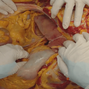
Case 22 Part 4
Abdominal survey — basic anatomy.
-
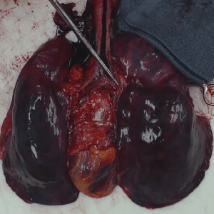
Case 22 Part 5
Heart and lung block — basic anatomy. Intrabronchial fluid (pulmonary edema).
-
Case 22 Part
Toxicology.
-

Case 23 – History
Recent laparoscopic surgery.
-
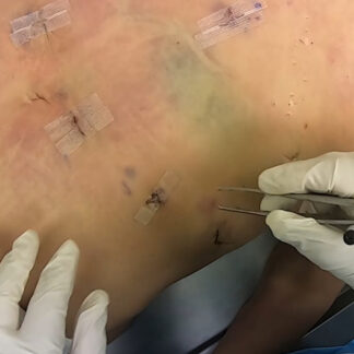
Case 23 Part 1
External exam.
-
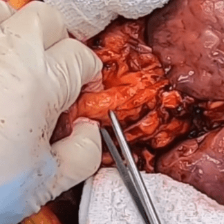
Case 23 Part 2
Isolation of airway.
-
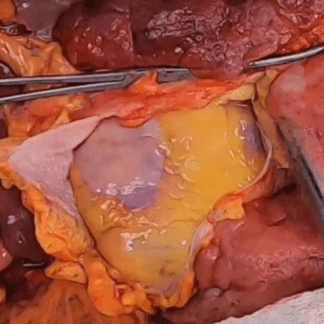
Case 23 Part 3
Assessment of chest.
-
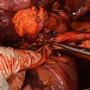
Case 23 Part 4
Exposure of esophageal hiatus.
-
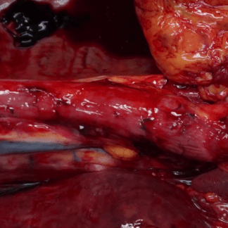
Case 23 Part 5
Roux-en-Y anatomy.
-

Case 24 – History
History of coronary artery bypass graft and recent back surgery.
-
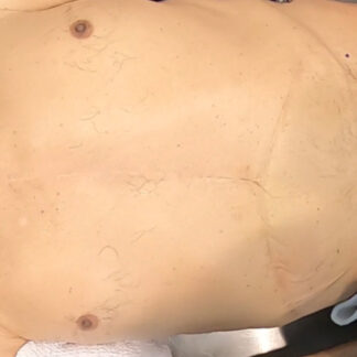
Case 24 Part 1
External exam.
-
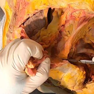
Case 24 Part 2
Y-shaped incision.
-
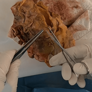
Case 24 Part 3
Coronary artery bypass graft surgery — ostial scarring with graft closure.
-

Case 25 – History
Motor vehicle accident and untreated hypertension.
-
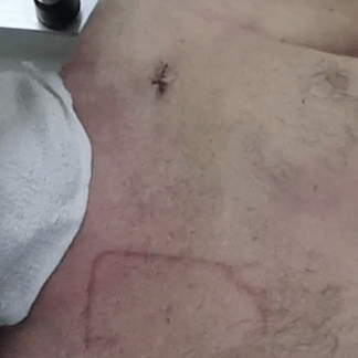
Case 25 Part 1
External exam. Low-speed motor vehicle accident.
-
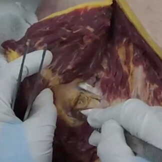
Case 25 Part 2
Y-shaped incision.
-
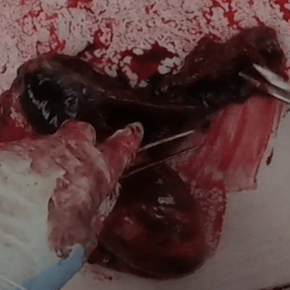
Case 25 Part 4
Aortic tear — hypertension
-

Case 26 – History
Dementia and nursing home fall.
-
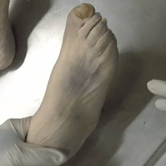
Case 26 Part 1
Contusions.
-
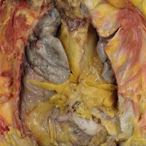
Case 26 Part 2
Chest wall contusions.
-
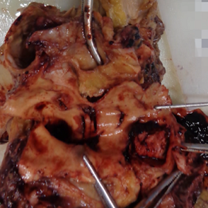
Case 26 Part 3
Pulmonary artery.
-
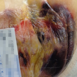
Case 26 Part 4
Scalp contusions.
-

Case 27 – History
Hypotension during dialysis.
-
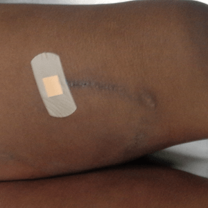
Case 27 Part 1
External exam. Multiple dialysis access surgeries.
-
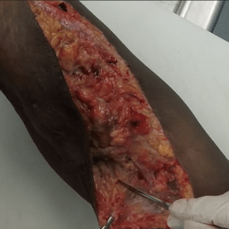
Case 27 Part 2
Left arm dissection. HeRO graft.
-
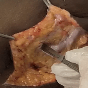
Case 27 Part 3
Left leg dissection. Dialysis graft.
-
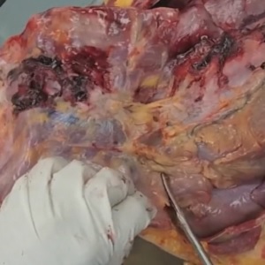
Case 27 Part 4
Removal of chest plate. Rib fractures from CPR.
-
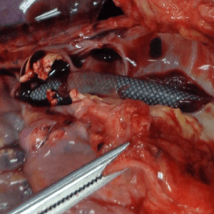
Case 27 Part 5
HeRO graft. Near-total obstruction of superior vena cava orifice.
-
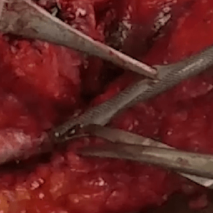
Case 27 Part 6
HeRO graft opened.
-
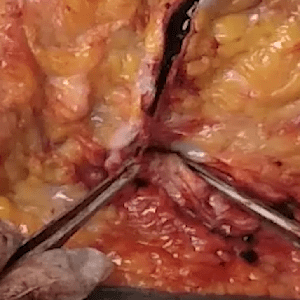
Case 27 Part 7
Left leg dialysis graft — opened.
-
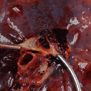
Case 27 Part 8
Chronic pulmonary embolism. Pulmonary infarcts.
-

Case 28 – History
Peripheral vascular disease and recent angioplasty.
-
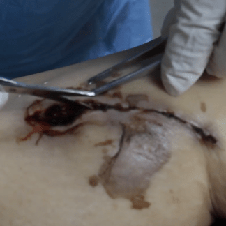
Case 28 Part 1
External exam. Reperfusion syndrome.
-
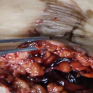
Case 28 Part 2
Compartment syndrome.
-
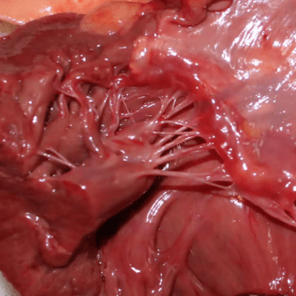
Case 28 Part 3
Acute myocardial infarction.
-

Case 28 Part 4
Pre- and post-operative angiograms. Echocardiogram.
-

Case 29 – History
Leg swelling.
-
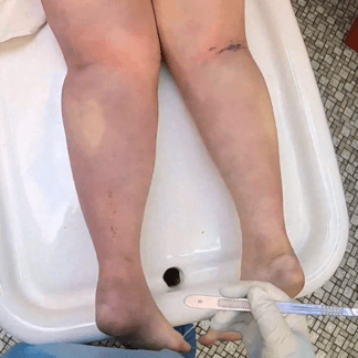
Case 29 Part 1
Unilateral leg swelling.
-
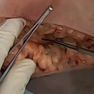
Case 29 Part 2
Lower extremity soft tissue edema.
-
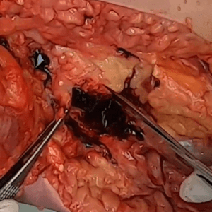
Case 29 Part 3
Deep venous thrombosis.
-
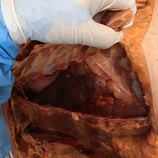
Case 29 Part 4
Acute pulmonary embolism.
-

Case 30 – History
Alcohol use disorder and patellar tendon repair.
-
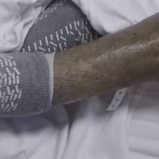
Case 30 Part 1
External exam.
-
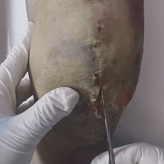
Case 30 Part 2
Patellar tendon tear. Surgical repair and repeat tear. In situ.
-
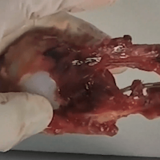
Case 30 Part 3
Patellar tendon tear. Surgical repair and repeat tear. Knee replacement.
-
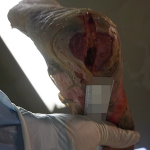
Case 30 Part 4
Heel decubitus ulcer
-
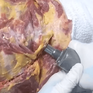
Case 30 Part 5
Y-shaped incision.
-
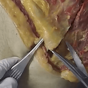
Case 30 Part 6
Chest plate removal.
-
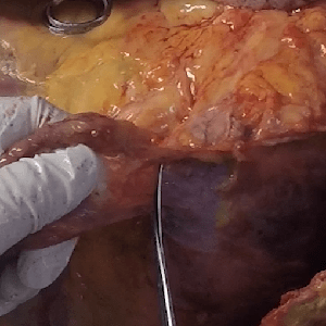
Case 30 Part 7
(Normal) thoracic post-surgical scarring after CABG.
-
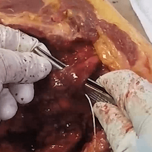
Case 30 Part 8
Basic anatomy — branches of aortic arch.
-
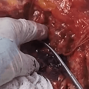
Case 30 Part 9
Removal of chest organs.
-
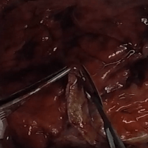
Case 30 Part 10
Saphenous vein graft dissection.
-
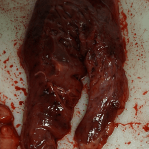
Case 30 Part 11
Old myocardial infarction.
-

Case 31 – History
Young male. Second autopsy.
-
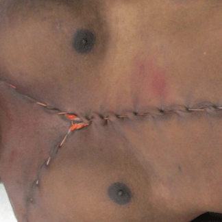
Case 31 Part 1
External exam. Lip laceration.
-
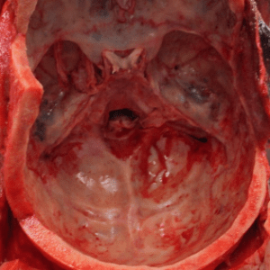
Case 31 Part 2
Defensive injuries. Scalp contusions. Petrous bone hemorrhage.
-

Case 32 – History
Elderly woman with dementia and postoperative lethargy.
-
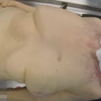
Case 32 Part 1
External exam.
-
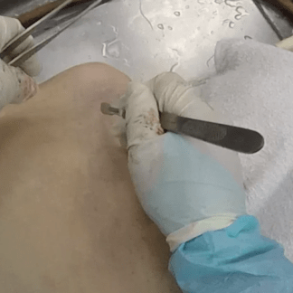
Case 32 Part 2
Y-shaped incision. Rib fractures.
-
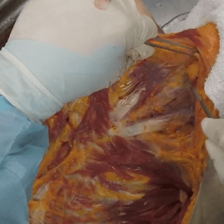
Case 32 Part 3
Review — basic neck anatomy.
-

Case 32 Part 4
Toxicology.
-

Case 33 – History
Middle-aged man with hypertension and chest pain.
-
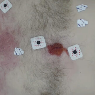
Case 33 Part 1
External exam. EKG leads.
-
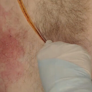
Case 33 Part 2
Y-shaped incision.
-
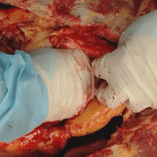
Case 33 Part 3
Tamponade.
-
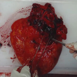
Case 33 Part 4
Aortic laceration/rupture.
-

Case 34 – History
Shortness of breath in an elderly woman with COPD.
-

Case 34 Part 1
External exam. Obesity. Cardiac catheterization femoral access site.
-
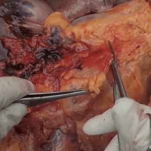
Case 34 Part 2
Pleural effusion. Chest wall and mediastinal hemorrhage. Cardiomegaly.
-
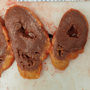
Case 34 Part 3
Severe aortic atherosclerosis. Coronary artery stent. Left ventricular hypertrophy.
-
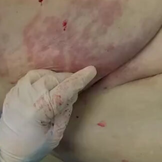
Case 34 Part 4
Exploration of catheter insertion site and abdominal wall.
-
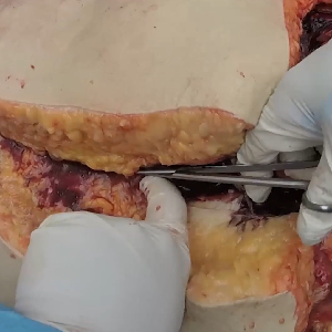
Case 34 Part 5
Exploration of abdominal wall and abdomen.
-
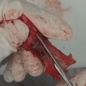
Case 34 Part 6
Iliofemoral artery with iatrogenic defect and stent.
-
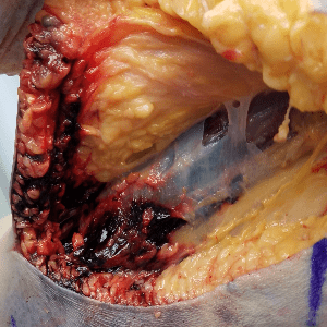
Case 34 Part 7
Assessment of right brachial artery access site.
-

Case 35 – History
Motor vehicle accident.
-
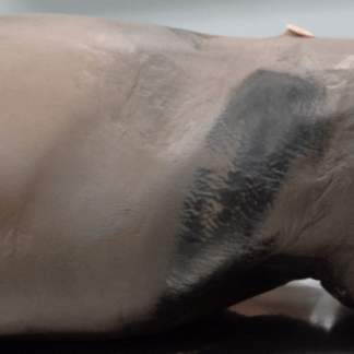
Case 35 Part 1
External exam. High-speed motor vehicle accident. Seat belt strap marks.
-
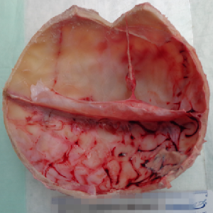
Case 35 Part 2
Skull and brain.
-
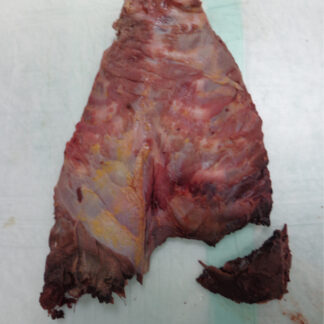
Case 35 Part 3
Initial survey chest and abdomen.
-
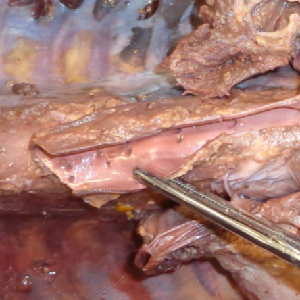
Case 35 Part 4
Severed spine.
-

Case 36 – History
Syncope.
-
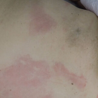
Case 36 Part 1
External exam. Livor mortis.
-
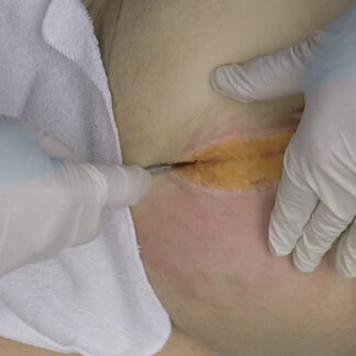
Case 36 Part 2
Hemoperitoneum.
-
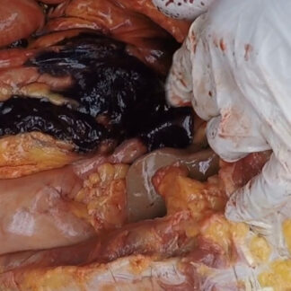
Case 36 Part 3
Intraperitoneal hematoma.
-

Case 37 – History
Alcohol use disorder and pacemaker replacement.
-
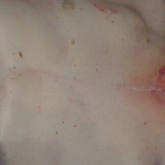
Case 37 Part 1
External exam. Disuse atrophy. Skin tear.
-
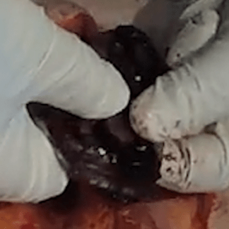
Case 37 Part 2
Pacemaker retrieval. Assessment of pacemaker surgical site.
-
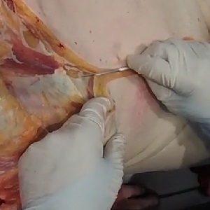
Case 37 Part 3
Abdominal wall anatomy. Ascites. Basic survey of abdominal organs.
-
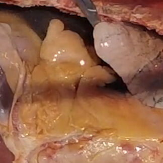
Case 37 Part 4
Removal of chest plate. Massive serous pleural effusions.
-
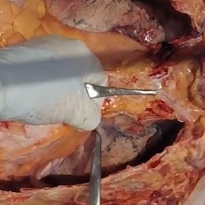
Case 37 Part 5
Post-surgical scarring. Checking for pulmonary embolism.
-
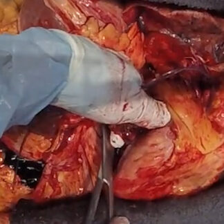
Case 37 Part 6
Removal of heart and lung block.
-
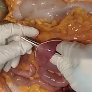
Case 37 Part 7
Removal of small intestine.
-
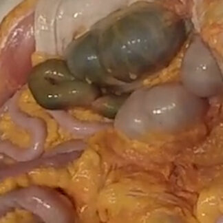
Case 37 Part 8
Removal of colon.
-
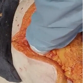
Case 37 Part 9
Removal of prostate.
-
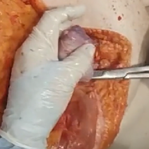
Case 37 Part 10
Removal of testes.
-
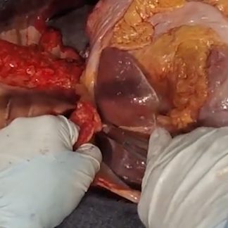
Case 37 Part 11
Removal of liver.
-
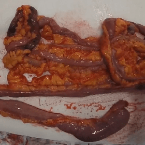
Case 37 Part 12
Opening the small intestine. Meckel’s diverticulum.
-

Case 37 Part 13
Opening the colon.
-
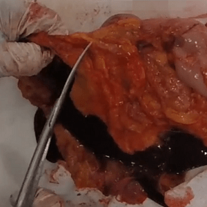
Case 37 Part 14
Duodenal varices.
-
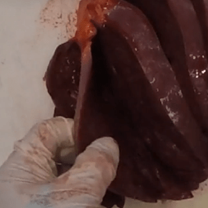
Case 37 Part 15
Liver. Gallbladder.
-

Case 38 – History
Lung mass.
-
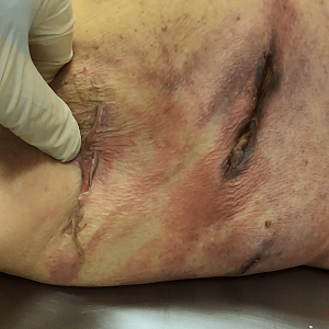
Case 38 Part 1
External exam. VATS procedure.
-
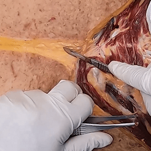
Case 38 Part 2
Y-shaped incision. CPR-related trauma.
-
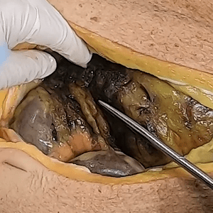
Case 38 Part 3
Pneumoperitoneum. Acute peritonitis.
-
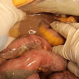
Case 38 Part 4
Acute peritonitis.
-
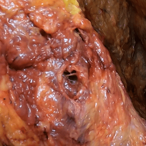
Case 38 Part 5
Assessment of VATS surgical site from skin to chest cavity.
-
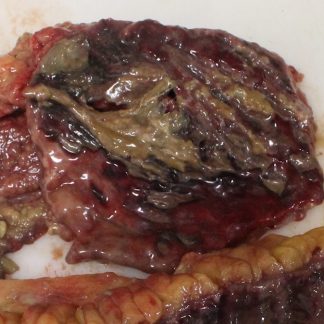
Case 38 Part 6
Crohn’s disease.
-
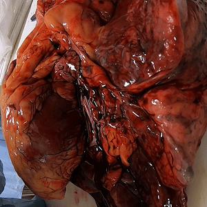
Case 38 Part 7
Heart and lung block.
-
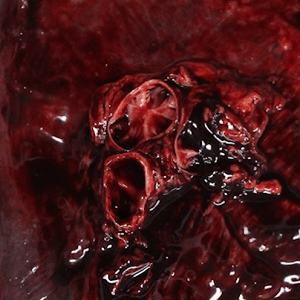
Case 38 Part 8
Pulmonary embolism.
-

Case 39 – History
Sudden collapse. History of alcohol use disorder (recovering).
-
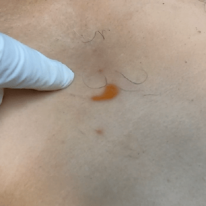
Case 39 Part 1
External exam.
-
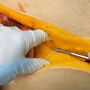
Case 39 Part 2
Hemoperitoneum.
-
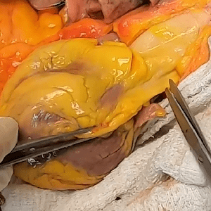
Case 39 Part 3
Assessing for pericardial fluid and pulmonary embolism.
-
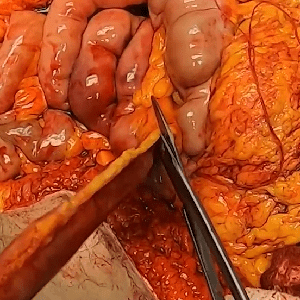
Case 39 Part 4
Removal of intestine.
-
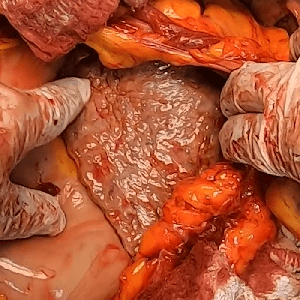
Case 39 Part 5
Cirrhosis.
-
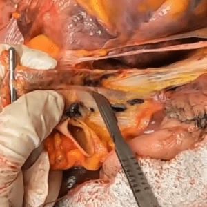
Case 39 Part 6
Thoracic adhesions.
-
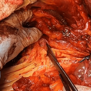
Case 39 Part 7
Abdominal aortic ostia.
-
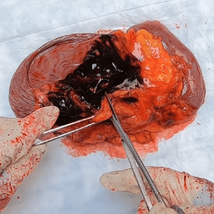
Case 39 Part 8
Splenomegaly. Splenic vein thrombosis.
-

Case 40 – History
History of smoking.
-
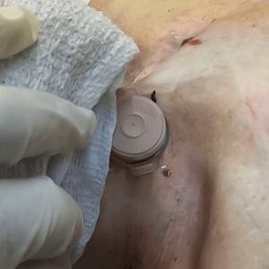
Case 40 Part 1
External exam. Tracheostomy with scarring. Muscle graft donor site. Gastrostomy tube.
-
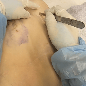
Case 40 Part 2
Y-shaped incision.
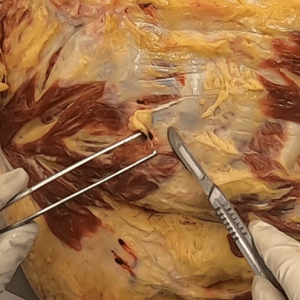
-
Case 40 Part 3
Assessment of biopsy needle track.
-
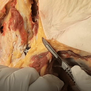
Case 40 Part 4
Chest plate removal.
-
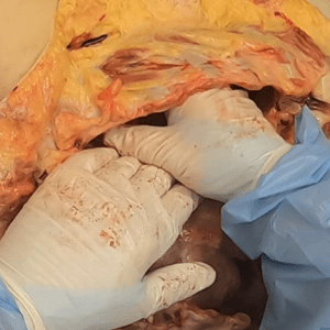
Case 40 Part 5
Hemothorax.
-
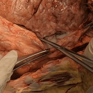
Case 40 Part 6
Intestinal autotransplant.
-
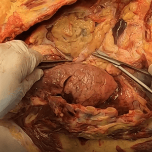
Case 40 Part 7
Thoracic postsurgical scarring and adhesions.
-
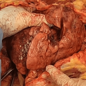
Case 40 Part 8
Right lung variant anatomy.
-
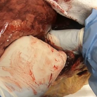
Case 40 Part 9
Removal of chest organs.
-
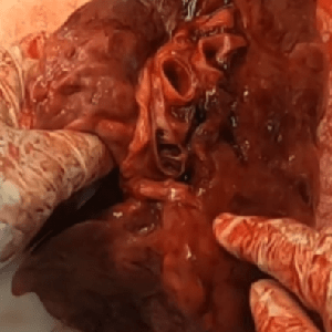
Case 40 Part 10
Basic hilar anatomy. Intrabronchial hemorrhage.
-
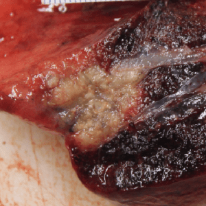
Case 40 Part 11
Lung cancer.
-

Case 41 – History
Recent weight gain.
-
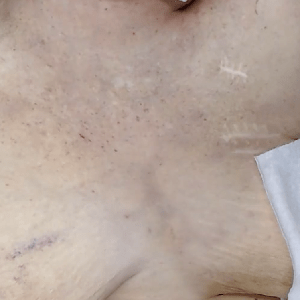
Case 41 Part 1
External exam.
-
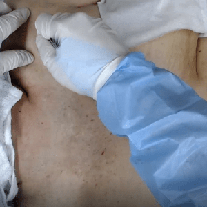
Case 41 Part 2
Anasarca. Tissue edema.
-
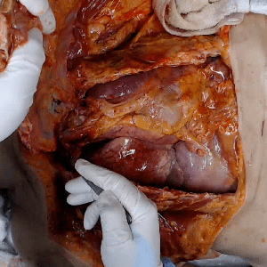
Case 41 Part 3
Anasarca. Pleural effusion.
-
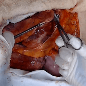
Case 41 Part 4
Uremic pericarditis.
-

Case 42 – History
Death during rehab.
-
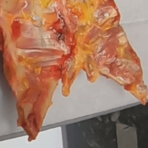
Case 42 Part 1
Bifid xiphoid process.
-
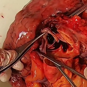
Case 42 Part 2
Acute pulmonary embolism.
-
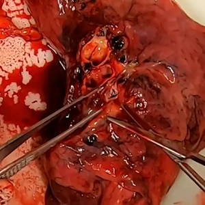
Case 42 Part 3
Subacute pulmonary embolism.
-
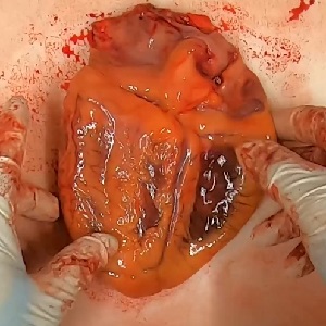
Case 42 Part 4
Heart — tour of basic external anatomy.
-
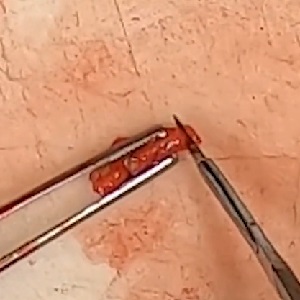
Case 42 Part 5
Severe coronary atherosclerosis.
-
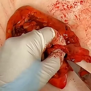
Case 42 Part 6
Heart — internal anatomy.
-

Case 43 – History
Mental status change followed by hypotension.
-
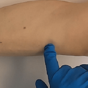
Case 43 Part 1
External exam. Head injury.
-
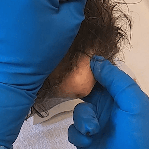
Case 43 Part 2
Skull exposure. Internal scalp assessment.
-
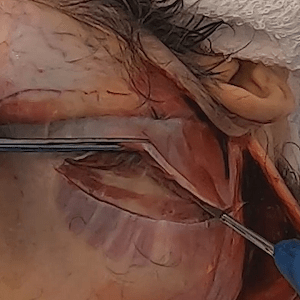
Case 43 Part 3
Coup-contrecoup injury. Brain in situ.
-
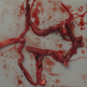
Case 43 Part 4
Coup-contrecoup injury. Brain removed.
-
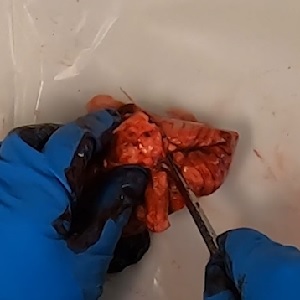
Case 43 Part 5
Duret hemorrhage.
-

Case 44 – History
Longstanding urinary incontinence and frequency.
-
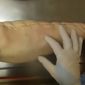
Case 44 Part 1
External exam. Skin, soft tissue and long bone donation.
-
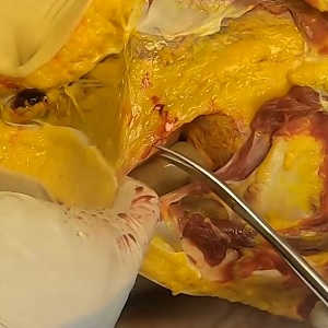
Case 44 Part 2
Hemoperitoneum.
-
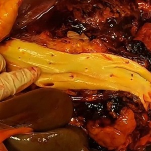
Case 44 Part 3
Assessment of aorta.
-
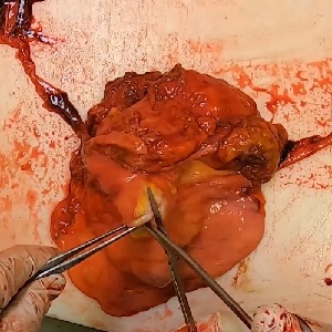
Case 44 Part 4
Massive prostatic hypertrophy.
-
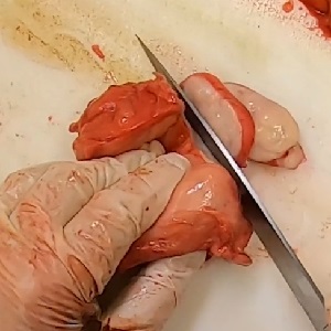
Case 44 Part 5
Prostate dissection.
-
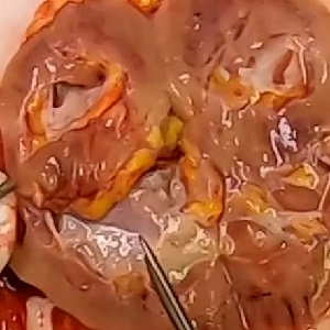
Case 44 Part 6
Hydroureter. Hydronephrosis.
-

Case 45 – History
Death during sleep.
-
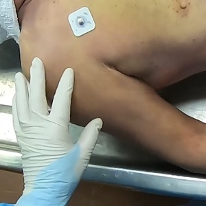
Case 45 Part 1
External exam.
-
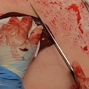
Case 45 Part 2
Pulmonary congestion and edema.
-
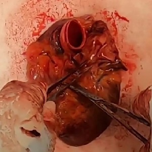
Case 45 Part 3
Left mainstem coronary artery blockage.
-

Case 46 – History
Pacemaker and chronic back pain.
-
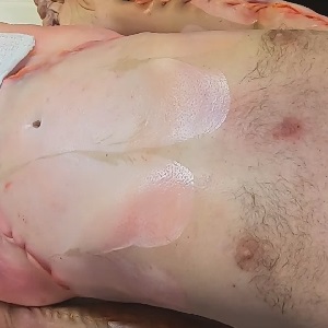
Case 46 Part 1
External exam. Skin donation.
-
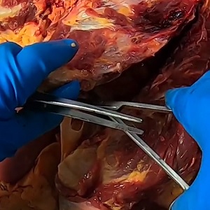
Case 46 Part 2
Subacute pacemaker lead perforation.
-
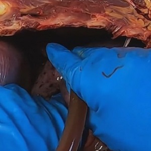
Case 46 Part 3
Empyema.
-
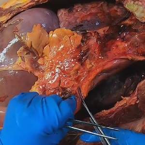
Case 46 Part 4
Severe acute pericarditis.
-

Case 47 – History
Middle-aged woman status post remote hysterectomy.
-
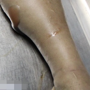
Case 47 Part 1
External exam. Multiple recent abdominal surgeries. Embalming. Decomposition.
-
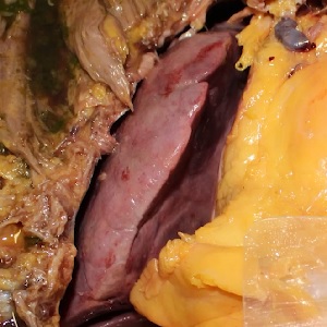
Case 47 Part 2
Massive cardiomegaly. Bilobed right lung.
-
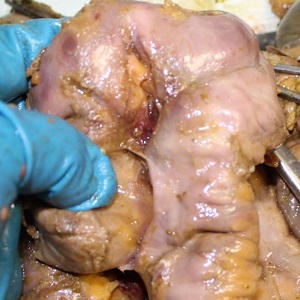
Case 47 Part 3
Dense intestinal adhesions. Gastric ulcer.
-

Case 48 – History
Death during dialysis.
-
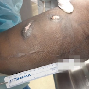
Case 48 Part 1
External exam. Arteriovenous fistula.
-
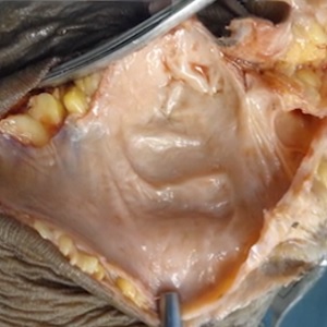
Case 48 Part 2
Dissection of arteriovenous fistula.
-
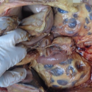
Case 48 Part 3
Polycystic kidney disease.
-

Case 49 – History
Recurrent liposarcoma.
-
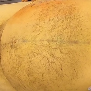
Case 49 Part 1
External exam. Jaundice.
-
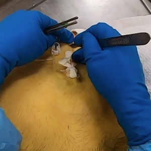
Case 49 Part 2
Y-shaped incision.
-

Case 49 Part 3
Post-surgical abdomen. Initial abdominal survey.
-

Case 50 – History
Death after coronary stenting.
-
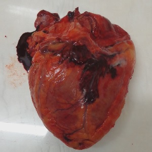
Case 50 Part 1
Cardiac amyloidosis.
-

Case 51 – History
Sudden death in rehab – in development.
-
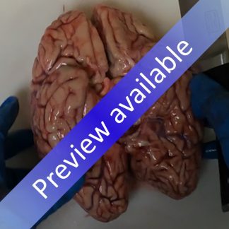
Case 51 Part 1
Brain dissection.
-

Case 52 – History
Sudden death after knee arthroscopy.
-
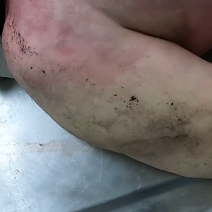
Case 52 Part 1
External exam.
-
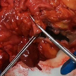
Case 52 Part 2
Gastric hemorrhage.
-
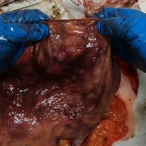
Case 52 Part 3
Gastric mucosa.
-
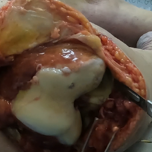
Case 52 Part 4
Knee surgery site.
-

Case 53 – History
Premature rupture of membranes.
-
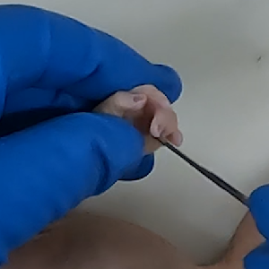
Case 53 Part 1
External exam.
-
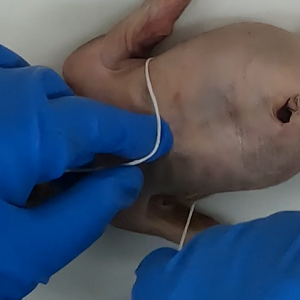
Case 53 Part 2
External exam – measurements.
-
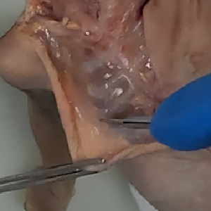
Case 53 Part 3
Y-shaped incision.
-
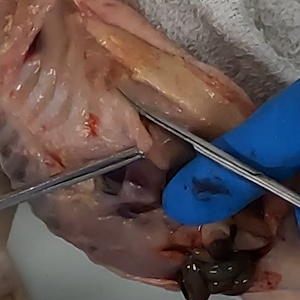
Case 53 Part 4
Removal of chest plate.
-
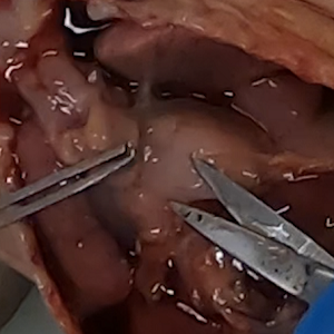
Case 53 Part 5
Survey of chest.
-
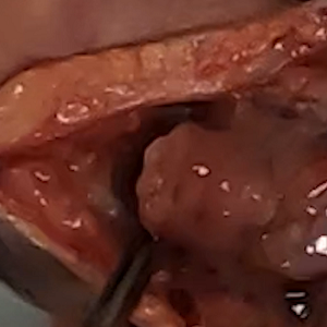
Case 53 Part 6
Isolation of larynx and trachea.
-
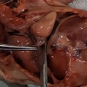
Case 53 Part 7
Removal of chest organs.
-
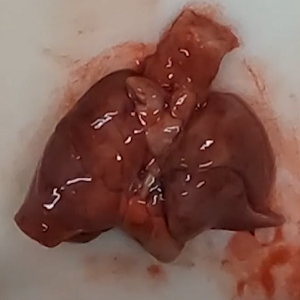
Case 53 Part 8
Survey of heart and lung block.
-
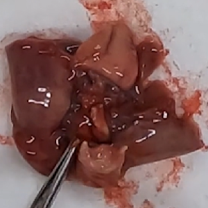
Case 53 Part 9
Airway and esophagus.
-
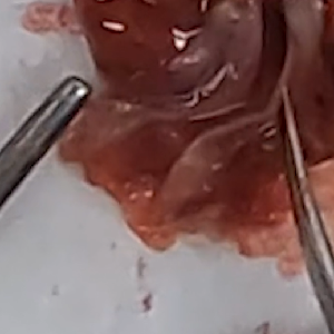
Case 53 Part 10
Opening airway.
-
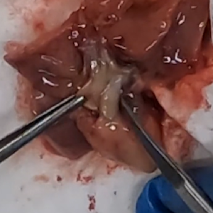
Case 53 Part 11
Ductus arteriosus.
-
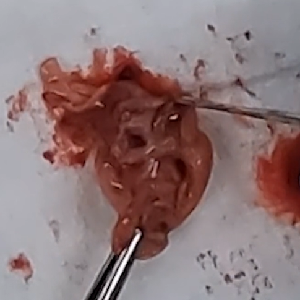
Case 53 Part 12
Foramen ovale.
-
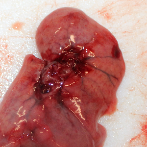
Case 53 Part 13
Lungs.
-
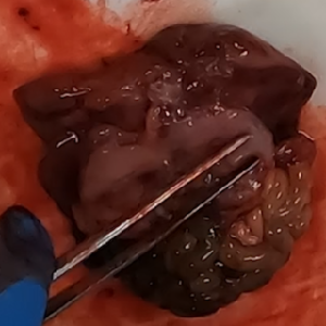
Case 53 Part 14
Abdominal organ block. Kidneys. Adrenals.
-
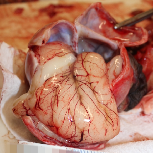
Case 53 Part 15
Abdominal organs. Skull. Brain.
