Library
Autopsy.Online
Desktop devices
recommended.
Autopsy.Online
How would you like to search the database?
By Visual Guide
By Case History
By Clinical Topic

Starting Tips

More ↓
Search the History for case information.
Starting Tips
Find suggestions to match your interest.
on larger devices.)
on tablet or larger devices.)
Back to top Showing all 320 results
-
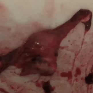
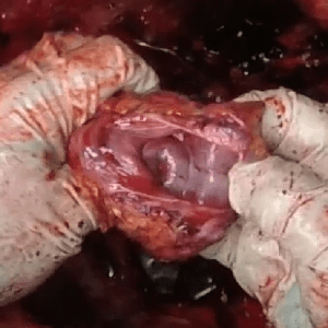
Case 50 Part 1
Cardiac amyloidosis
-
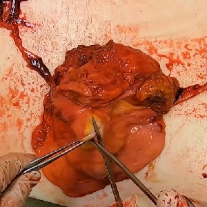
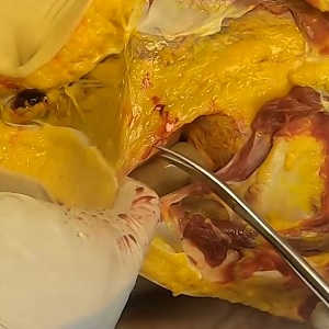
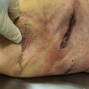
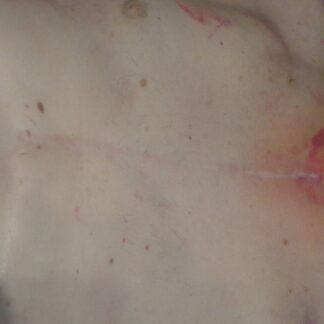
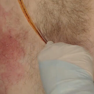
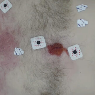
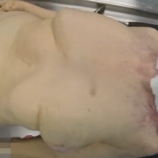
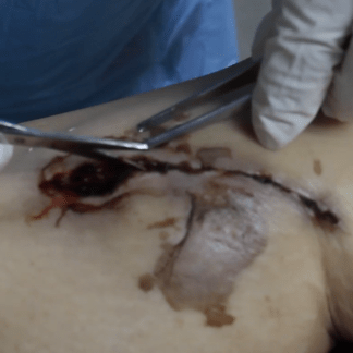
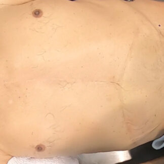
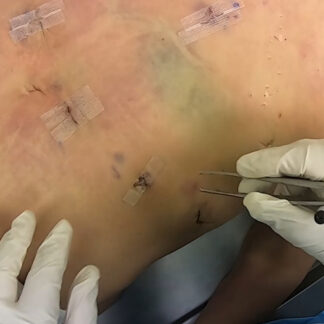
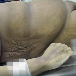
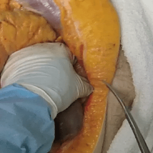
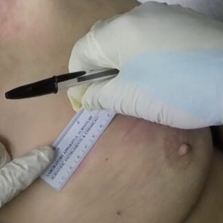
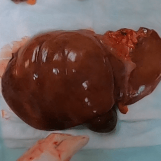
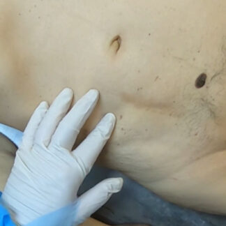
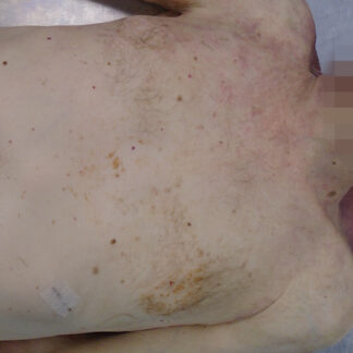
Case 49 Part 1
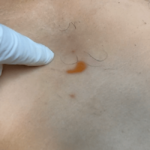
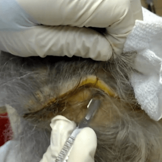
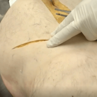
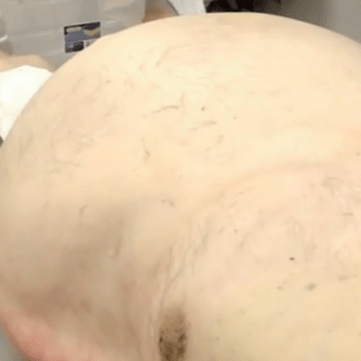
External exam. Jaundice.
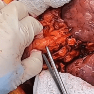
-

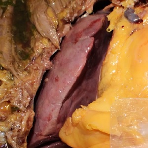
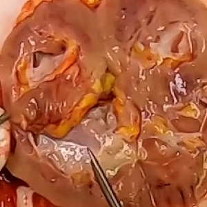
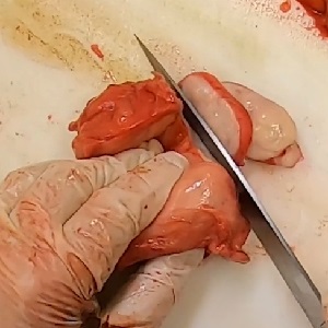
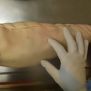
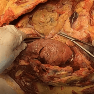
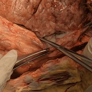
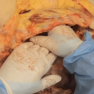
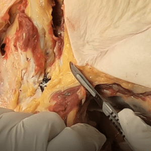
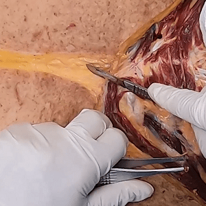
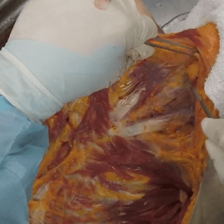
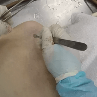
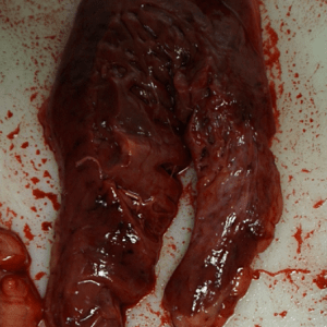
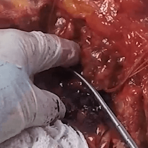
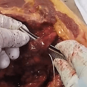
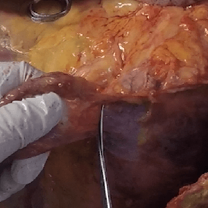
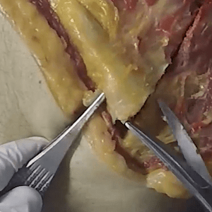
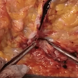
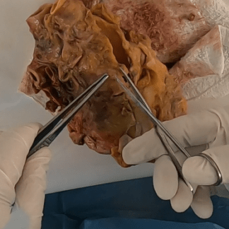
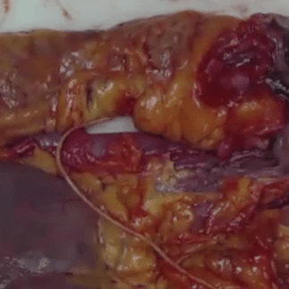
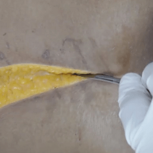
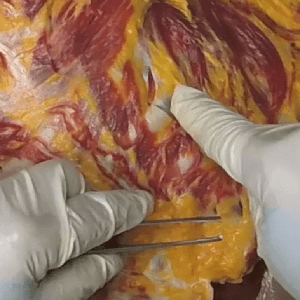
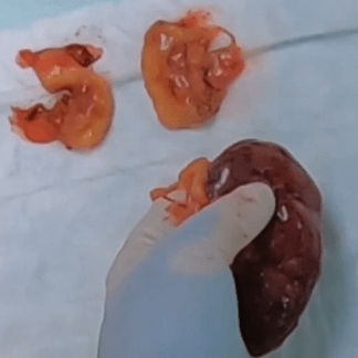
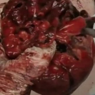
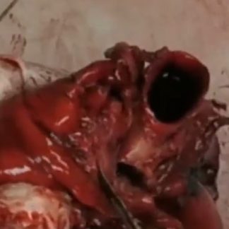
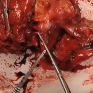
Case 49 – History
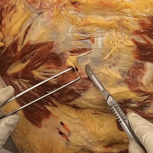
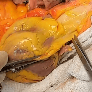
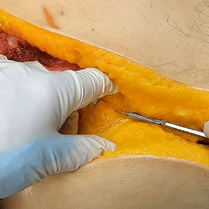
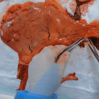
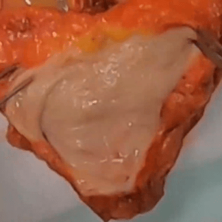
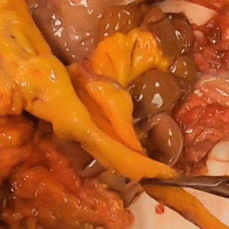
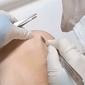
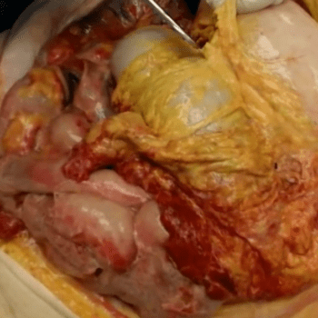
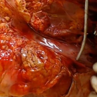
Recurrent liposarcoma.
-
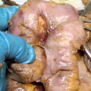
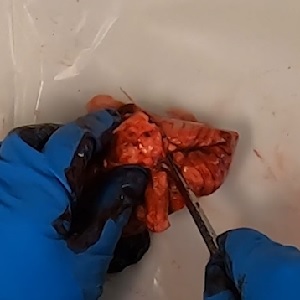
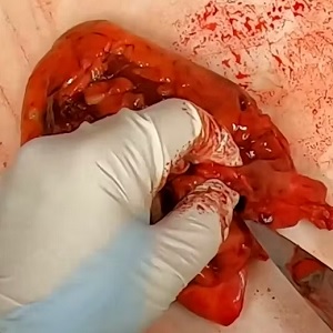
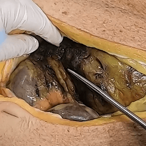
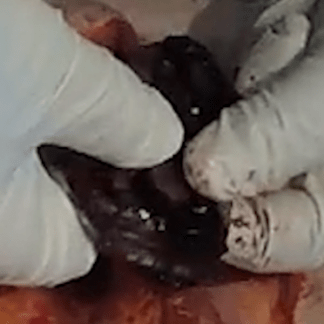
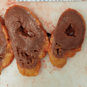
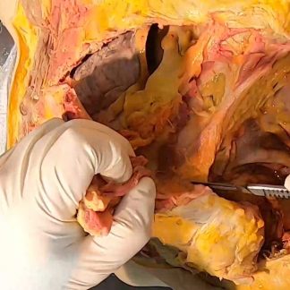
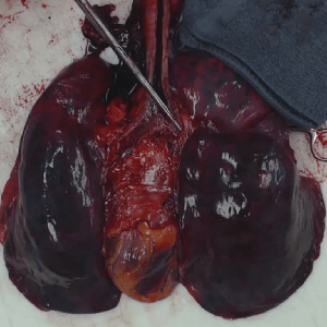
Case 48 Part 3
Polycystic kidney disease.
-
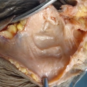
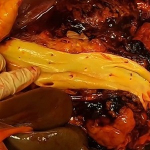
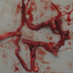
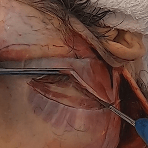
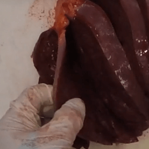
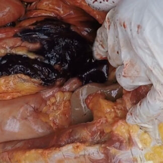
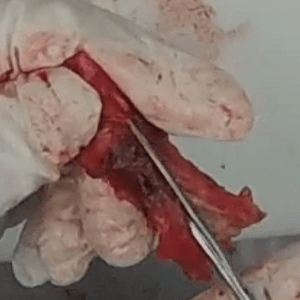
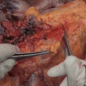
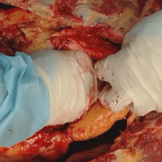
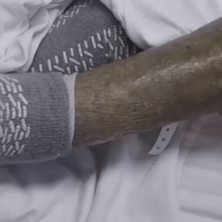
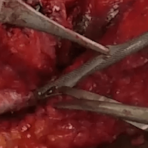
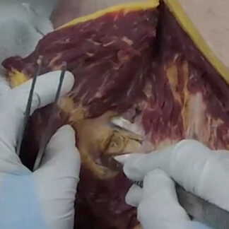
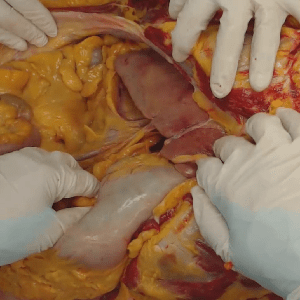
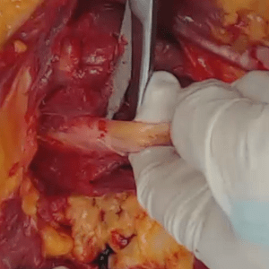
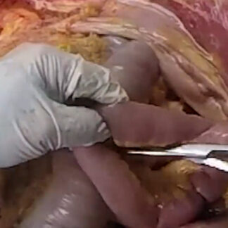
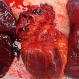
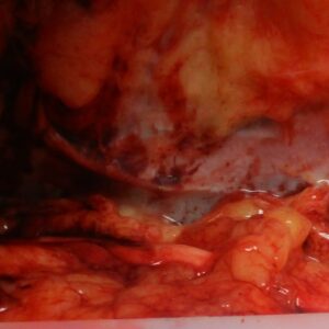
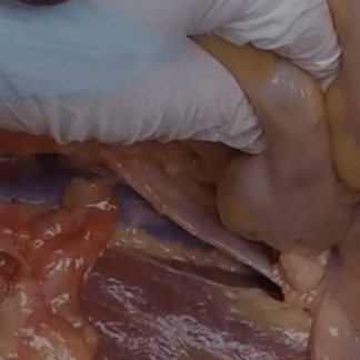
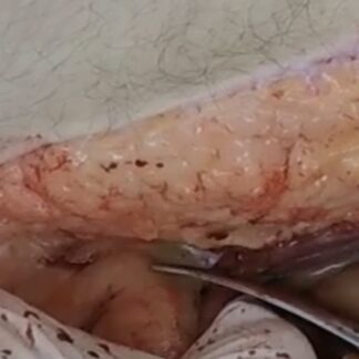
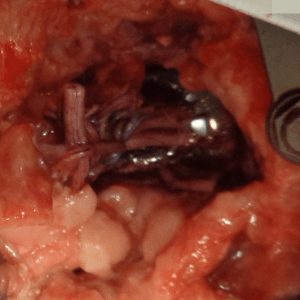
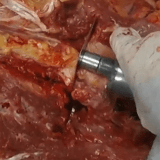
Case 48 Part 2
Dissection of arteriovenous fistula.
-
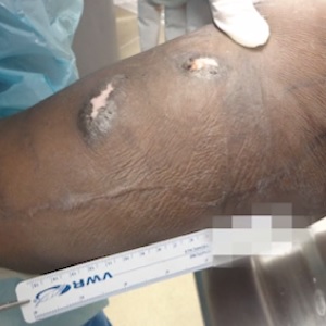
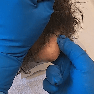
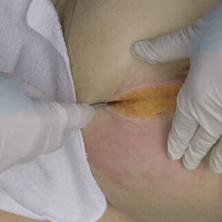
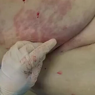
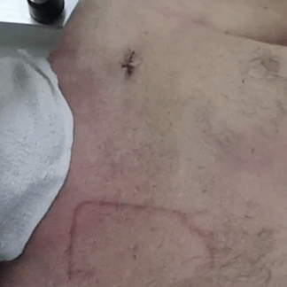
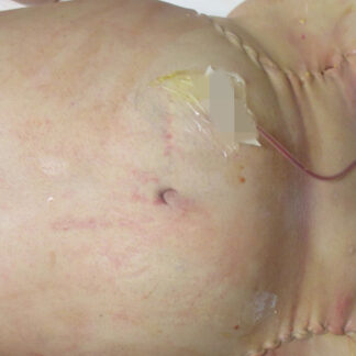
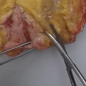
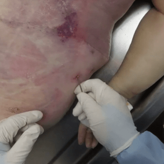
Case 48 Part 1
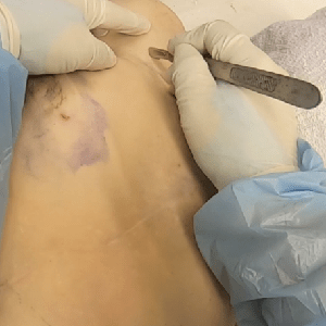
External exam. Arteriovenous fistula.
-
Case 48 – History
Death during dialysis.
-
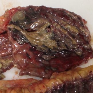
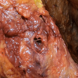
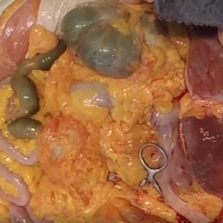
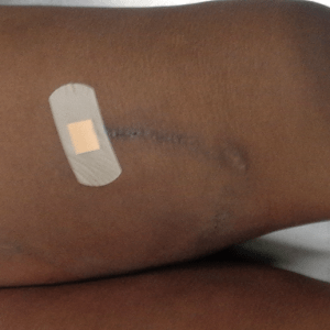
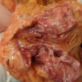
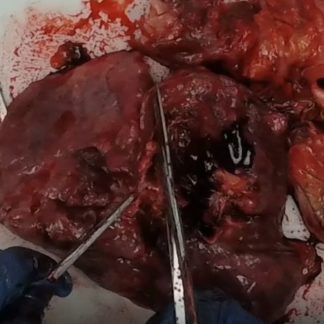
Case 47 Part 3
Dense intestinal adhesions. Gastric ulcer.
-
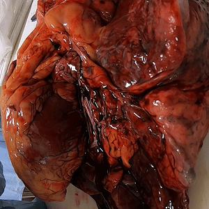
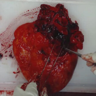
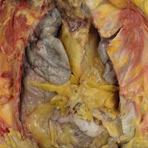
Case 47 Part 2
Massive cardiomegaly. Bilobed right lung.
-
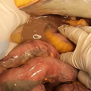
Case 47 Part 1
External exam. Multiple recent abdominal surgeries. Embalming. Decomposition.
-
Case 47 – History
Middle-aged woman status post remote hysterectomy.
-
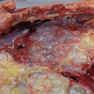
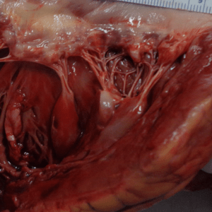
Case 46 Part 4
Severe acute pericarditis.
-
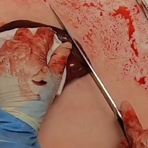
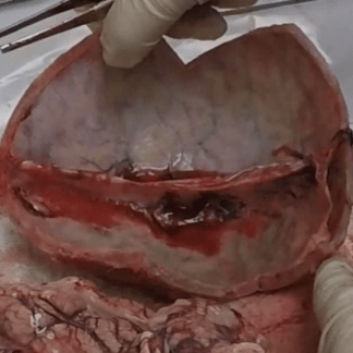
Case 46 Part 3
Empyema.
-
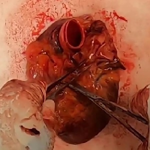
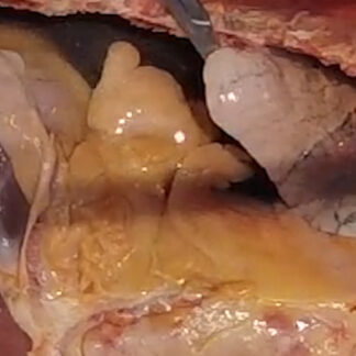
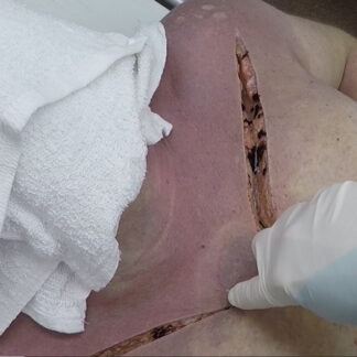
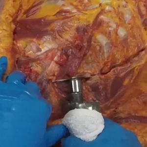
Case 46 Part 2
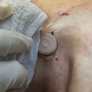
Subacute pacemaker lead perforation.
-
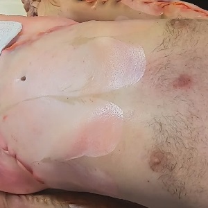
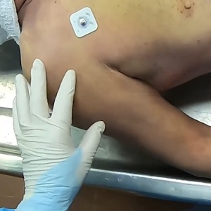
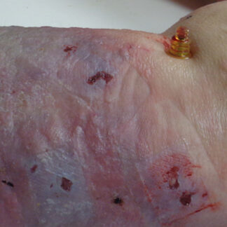
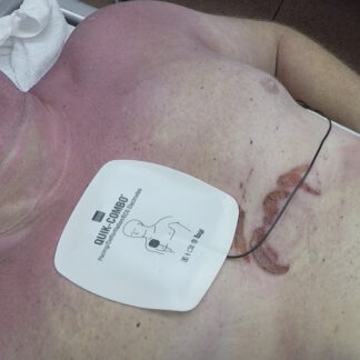
Case 46 Part 1
External exam. Skin donation.
-
Case 46 – History
Pacemaker and chronic back pain.
-
Case 45 Part 3
Left mainstem coronary artery blockage.
-
Case 45 Part 2
Pulmonary congestion and edema.
-
Case 45 Part 1
External exam.
-
Case 45 – History
Death during sleep.
-
Case 44 Part 6
Hydroureter. Hydronephrosis.
-
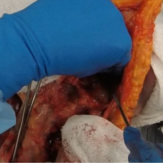
Case 44 Part 5
Prostate dissection.
-
Case 44 Part 4
Massive prostatic hypertrophy.
-
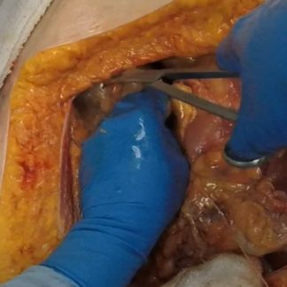
Case 44 Part 3
Assessment of aorta.
-
Case 44 Part 2
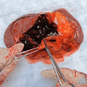
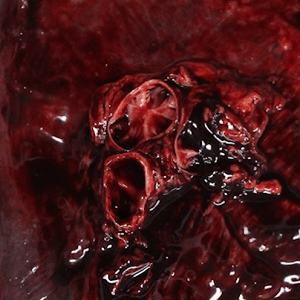
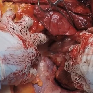
Hemoperitoneum.
-
Case 44 Part 1
External exam. Skin, soft tissue and long bone donation.
-
Case 44 – History
Longstanding urinary incontinence and frequency.
-
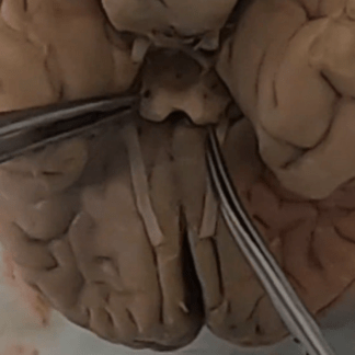
Case 43 Part 5
Duret hemorrhage.
-
Case 43 Part 4
Coup-contrecoup injury. Brain removed.
-
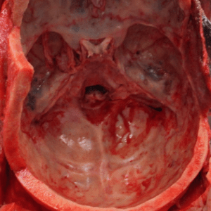
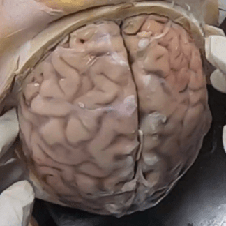
Case 43 Part 3
Coup-contrecoup injury. Brain in situ.
-
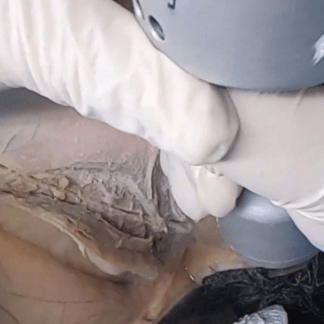
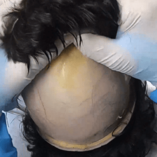
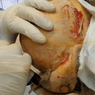
Case 43 Part 2
Skull exposure. Internal scalp assessment.
-
Case 43 Part 1
External exam. Head injury.
-
Case 43 – History
Mental status change followed by hypotension.
-
Case 42 Part 6
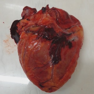
Heart — internal anatomy.
-
Case 42 Part 5
Severe coronary atherosclerosis.
-
Case 42 Part 4
Heart — tour of basic external anatomy.
-
Case 42 Part 3
Subacute pulmonary embolism.
-
Case 42 Part 2
Acute pulmonary embolism.
-
Case 42 Part 1
Bifid xiphoid process.
-
Case 42 – History
Death during rehab.
-
Case 41 Part 4
Uremic pericarditis.
-
Case 41 Part 3
Anasarca. Pleural effusion.
-
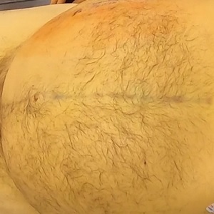
Case 41 Part 2
Anasarca. Tissue edema.
-

Case 41 Part 1
External exam.
-
Case 41 – History
Recent weight gain.
-
Case 40 Part 11
Lung cancer.
-
Case 40 Part 10
Basic hilar anatomy. Intrabronchial hemorrhage.
-
Case 40 Part 9
Removal of chest organs.
-
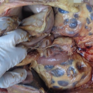
Case 40 Part 8
Right lung variant anatomy.
-
Case 40 Part 7
Thoracic postsurgical scarring and adhesions.
-
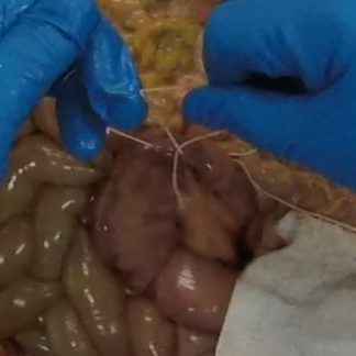
Case 40 Part 6
Intestinal autotransplant.
-
Case 40 Part 5
Hemothorax.
-
Case 40 Part 4
Chest plate removal.
-
Case 40 Part 3
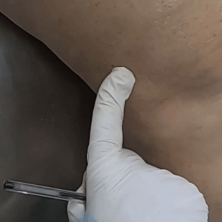
Assessment of biopsy needle track.
-
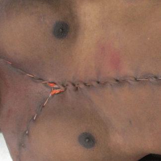
Case 40 Part 2
Y-shaped incision.
-
Case 40 Part 1
External exam. Tracheostomy with scarring. Muscle graft donor site. Gastrostomy tube.
-
Case 40 – History
History of smoking.
-
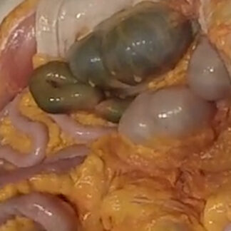
Case 39 Part 8
Splenomegaly. Splenic vein thrombosis.
-
Case 39 Part 7
Abdominal aortic ostia.
-
Case 39 Part 6
Thoracic adhesions.
-
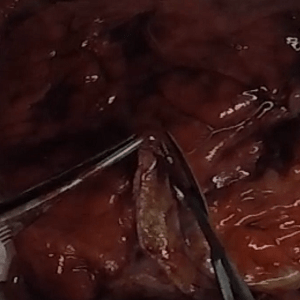
Case 39 Part 5
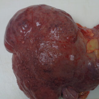
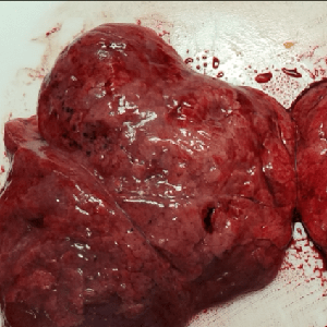
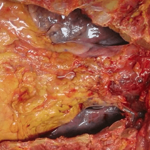
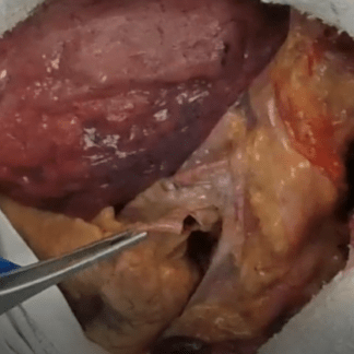
Cirrhosis.
-
Case 39 Part 4
Removal of intestine.
-
Case 39 Part 3
Assessing for pericardial fluid and pulmonary embolism.
-
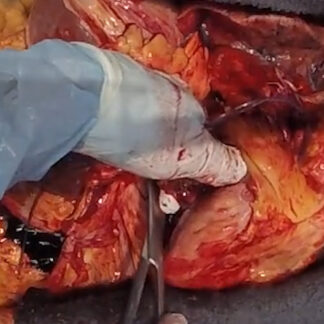
Case 39 Part 2
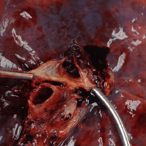
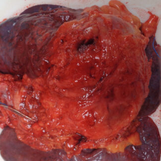
Hemoperitoneum.
-
Case 39 Part 1
External exam.
-
Case 39 – History
Sudden collapse. History of alcohol use disorder (recovering).
-
Case 38 Part 8
Pulmonary embolism.
-
Case 38 Part 7
Heart and lung block.
-
Case 38 Part 6
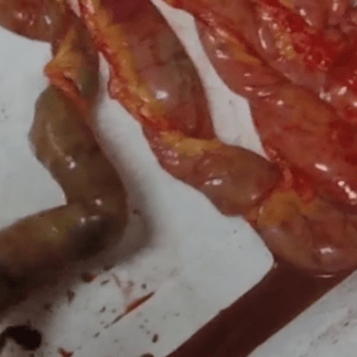
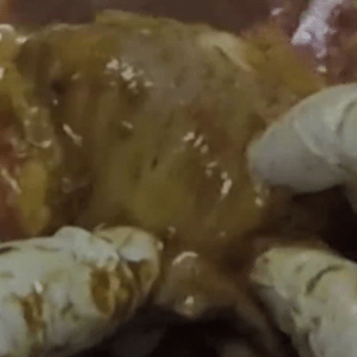
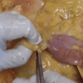
Crohn’s disease.
-
Case 38 Part 5
Assessment of VATS surgical site from skin to chest cavity.
-
Case 38 Part 4
Acute peritonitis.
-
Case 38 Part 3
Pneumoperitoneum. Acute peritonitis.
-
Case 38 Part 2
Y-shaped incision. CPR-related trauma.
-
Case 38 Part 1
External exam. VATS procedure.
-
Case 38 – History
Lung mass.
-
Case 37 Part 11
Liver. Gallbladder.
-
Case 37 Part 10
Duodenal varices.
-
Case 37 Part 9
Meckel’s diverticulum.
-
Case 37 Part 8
Removal of abdominal organs.
-
Case 37 Part 7
Bladder, prostate, testes — removal and basic anatomy.
-
Case 37 Part 6
Removal of small intestine.
-
Case 37 Part 5
Removal of chest organs. Scoliosis.
-
Case 37 Part 4
Removal of chest plate. Massive serous pleural effusions.
-
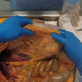
Case 37 Part 3
Ascites. Basic survey of abdominal organs.
-
Case 37 Part 2
Pacemaker retrieval. Assessment of pacemaker surgical site.
-
Case 37 Part 1
External exam. Disuse atrophy. Skin tear.
-
Case 37 – History
Alcohol use disorder and pacemaker replacement.
-
Case 36 Part 3
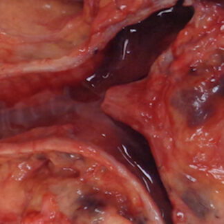
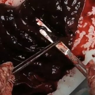
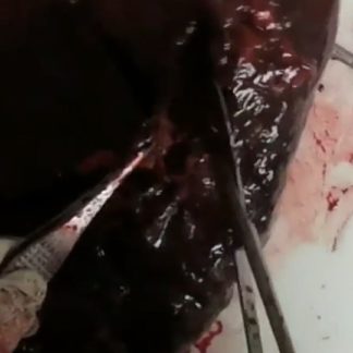
Intraperitoneal hematoma.
-
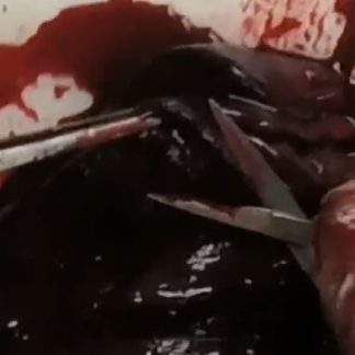
Case 36 Part 2
Hemoperitoneum.
-
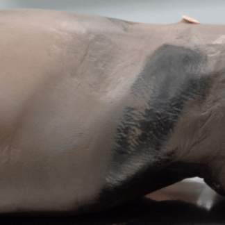
Case 36 Part 1
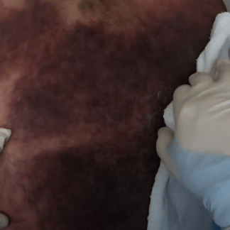
External exam. Livor mortis.
-
Case 36 – History
Syncope.
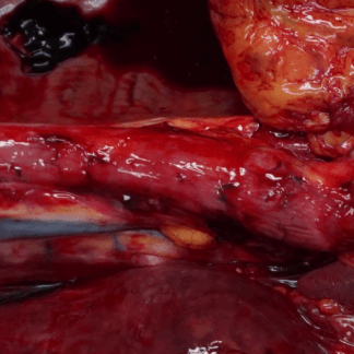
-
Case 35 Part 2
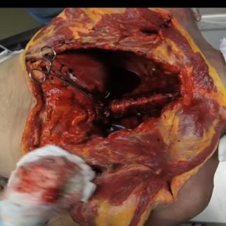
Severe internal trauma. Severed spine. Lacerated aorta and inferior vena cava.
-
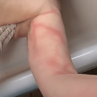
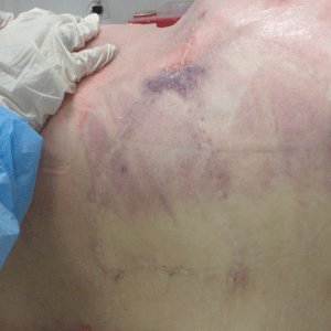
Case 35 Part 1
External exam. High-speed motor vehicle accident. Seat belt strap marks.
-
Case 35 – History
Motor vehicle accident.
-
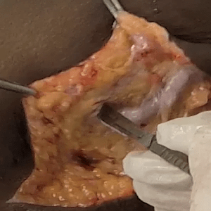
Case 34 Part 6
Assessment of right brachial artery access site.
-
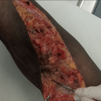
Case 34 Part 5
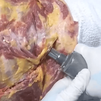
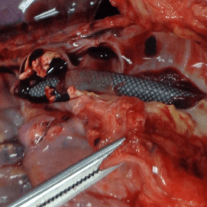
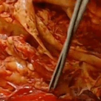
Iliofemoral artery with iatrogenic defect and stent.
-
Case 34 Part 4
Mental status change and massive hemorrhage after cardiac cath.
-
Case 34 Part 3
Severe aortic atherosclerosis. Coronary artery stent. Left ventricular hypertrophy.
-
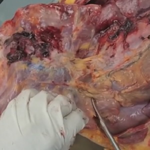
Case 34 Part 2
Pleural effusion. Chest wall and mediastinal hemorrhage. Cardiomegaly.
-
Case 34 Part 1
External exam. Obesity. Cardiac catheterization femoral access site.
-
Case 34 – History
Shortness of breath in an elderly woman with COPD.
-
Case 33 Part 4
Aortic laceration/rupture.
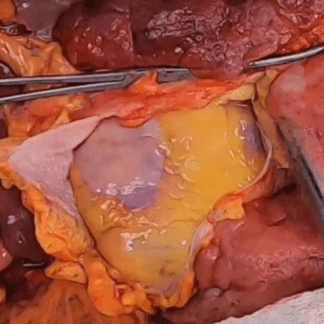
-
Case 33 Part 3
Tamponade.
-
Case 33 Part 2
Y-shaped incision.
-
Case 33 Part 1
External exam. EKG leads.
-
Case 33 – History
Middle-aged man with hypertension and chest pain.
-

Case 32 Part 4
Toxicology.
-
Case 32 Part 3
Review — basic neck anatomy.
-
Case 32 Part 2
Y-shaped incision. Rib fractures.
-
Case 32 Part 1
External exam.
-
Case 32 – History
Elderly woman with dementia and postoperative lethargy.
-
Case 31 Part 2
Defensive injuries. Scalp contusions. Petrous bone hemorrhage.
-
Case 31 Part 1
External exam. Lip laceration.
-
Case 31 – History
Young male. Second autopsy.
-
Case 30 Part 11
Old myocardial infarction.
-
Case 30 Part 10
Saphenous vein graft dissection.
-
Case 30 Part 9
Removal of chest organs.
-
Case 30 Part 8
Basic anatomy — branches of aortic arch.
-
Case 30 Part 7
(Normal) thoracic post-surgical scarring after CABG.
-
Case 30 Part 6
Chest plate removal.
-
Case 30 Part 5
Y-shaped incision.
-
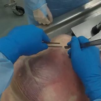
Case 30 Part 4
Heel decubitus ulcer
-
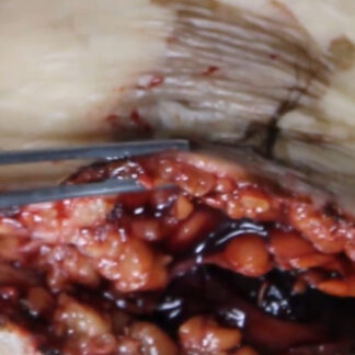
Case 30 Part 3
Patellar tendon tear. Surgical repair and repeat tear. Knee replacement.
-
Case 30 Part 2
Patellar tendon tear. Surgical repair and repeat tear. In situ.
-
Case 30 Part 1
External exam.
-
Case 30 – History
Alcohol use disorder and patellar tendon repair.
-
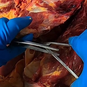
Case 29 Part 4
Acute pulmonary embolism.
-
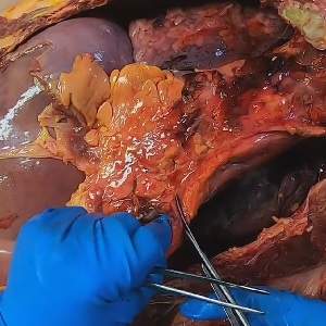
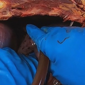
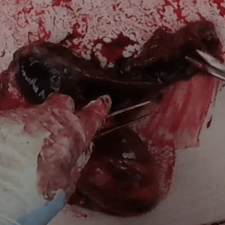
Case 29 Part 3
Deep venous thrombosis.
-
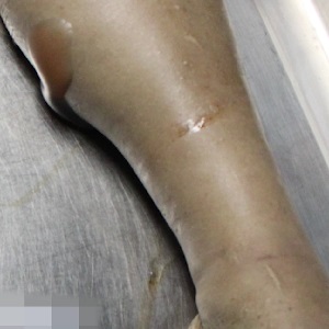
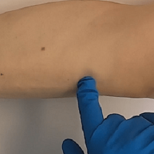
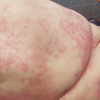
Case 29 Part 2
Lower extremity soft tissue edema.
-
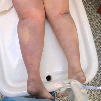
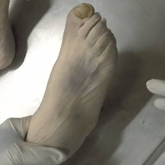
Case 29 Part 1
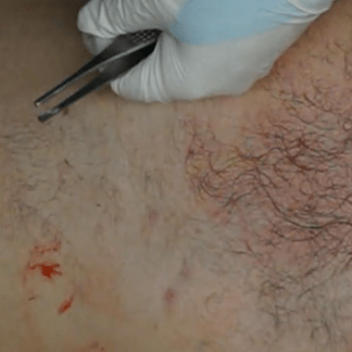
Unilateral leg swelling.
-
Case 29 – History
Leg swelling.
-
Case 28 Part 4
Pre- and post-operative angiograms. Echocardiogram.
-
Case 28 Part 3
Acute myocardial infarction.
-
Case 28 Part 2
Compartment syndrome.
-
Case 28 Part 1
External exam. Reperfusion syndrome.
-
Case 28 – History
Peripheral vascular disease and recent angioplasty.
-
Case 27 Part 8
Chronic pulmonary embolism. Pulmonary infarcts.
-
Case 27 Part 7
Left leg dialysis graft — opened.
-
Case 27 Part 6
HeRO graft opened.
-
Case 27 Part 5
HeRO graft. Near-total obstruction of superior vena cava orifice.
-
Case 27 Part 4
Removal of chest plate. Rib fractures from CPR.
-
Case 27 Part 3
Left leg dissection. Dialysis graft.
-
Case 27 Part 2
Left arm dissection. HeRO graft.
-
Case 27 Part 1
External exam. Multiple dialysis access surgeries.
-
Case 27 – History
Hypotension during dialysis.
-
Case 26 Part 2
Scalp and chest wall contusions. Pulmonary embolism.
-
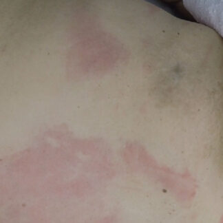
Case 26 Part 1
Contusions.
-
Case 26 – History
Dementia and nursing home fall.
-
Case 25 Part 3
Aortic tear — hypertension
-
Case 25 Part 2
Y-shaped incision.
-
Case 25 Part 1
External exam. Low-speed motor vehicle accident.
-
Case 25 – History
Motor vehicle accident and untreated hypertension.
-
Case 24 Part 3
Coronary artery bypass graft surgery — ostial scarring with graft closure.
-
Case 24 Part 2
Y-shaped incision.
-
Case 24 Part 1
External exam.
-
Case 24 – History
History of coronary artery bypass graft and recent back surgery.
-
Case 23 Part 4
Roux-en-Y anatomy.
-
Case 23 Part 3
Removal of chest organs.
-
Case 23 Part 2
Assessment of chest organs.
-
Case 23 Part 1
External exam.
-
Case 23 – History
Recent laparoscopic surgery.
-

Case 22 Part 7
Toxicology.
-
Case 22 Part 6
Abdominal organ dissection — basic anatomy.
-
Case 22 Part 5
Heart and lung block — basic anatomy. Intrabronchial fluid (pulmonary edema).
-
Case 22 Part 4
Abdominal survey — basic anatomy.
-
Case 22 Part 3
Neck dissection — basic anatomy.
-
Case 22 Part 2
Y-shaped incision.
-
Case 22 Part 1
External exam.
-
Case 22 – History
Middle-aged woman with psychiatric history.
-
Case 21 Part 5
Uterus dissection — basic anatomy.
-
Case 21 Part 4
Uterus — basic external anatomy.
-
Case 21 Part 3
Abdominal survey.
-
Case 21 Part 2
Removal of chest plate.
-
Case 21 Part 1
External exam.
-
Case 21 – History
History of breast cancer and alcohol use.
-
Case 20 Part 7
Old cavitary cerebral infarction.
-
Case 20 Part 6
Subcarinal lymph nodes. Duodenal, gastric and esophageal mucosa. Ampulla of Vater. Basic anatomy.
-
Case 20 Part 5
Ureter. Bladder. Prostate. Rectum. Basic anatomy.
-
Case 20 Part 4
Gastrointestinal tract. Ureter. Bladder.
-
Case 20 Part 3
Liver. Gallbladder. Basic anatomy.
-
Case 20 Part 2
Abdominal aorta – main branches. Inferior vena cava. Gerota’s fascia. Kidney. Ureter. Adrenals. Basic anatomy.
-
Case 20 Part 1
External exam.
-
Case 20 – History
Elderly man with stroke.
-
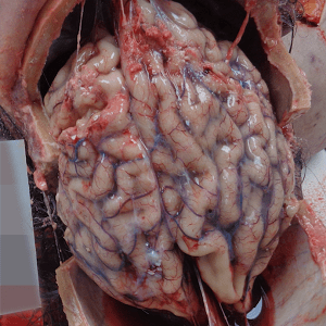
Case 19 Part 6
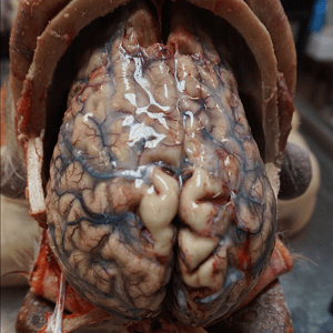
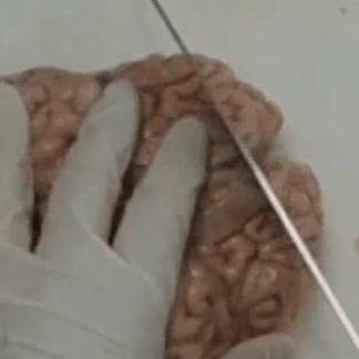
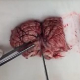
Brain — detailed internal anatomy.
-
Case 19 Part 5
Brain — detailed external anatomy.
-
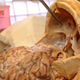
Case 19 Part 4
Brain tumor.
-
Case 19 Part 3
Brain exposure.
-
Case 19 Part 2
Skull exposure.
-
Case 19 Part 1
External exam.
-
Case 19 – History
Hypertension, dizziness and sudden death.
-
Case 18 Part 3
Brain exposure.
-
Case 18 Part 2
Skull exposure.
-
Case 18 Part 1
External exam.
-
Case 18 – History
Decomposition. History of clipped cerebral aneurysm.
-
Case 17 Part 6
Endotracheal tube.
-
Case 17 Part 5
Heart — basic anatomy. Coronary artery blockage.
-
Case 17 Part 4
Chest evaluation. Pleural effusion.
-
Case 17 Part 3
Rib fractures.
-
Case 17 Part 2
Y-shaped incision.
-
Case 17 Part 1
External exam. Early decomposition.
-
Case 17 – History
Sudden death.
-
Case 16 Part 3
Brain and base of skull — basic anatomy.
-
Case 16 Part 2
Brain exposure.
-
Case 16 Part 1
Skull exposure.
-
Case 16 – History
Dementia and kidney failure.
-
Case 15 Part 3
Toxicology.
-
Case 15 Part 2
Distal forearm musculature — detailed anatomy.
-
Case 15 Part 1
External exam.
-
Case 15 – History
Chronic pain and sudden death.
-
Case 14 Part 9
Heart assessment.
-
Case 14 Part 8
Lung assessment.
-
Case 14 Part 7
Lung assessment. Pulmonary edema.
-
Case 14 Part 6
Heart and lung block.
-
Case 14 Part 5
Diaphragm — basic anatomy.
-
Case 14 Part 4
Chest assessment.
-
Case 14 Part 3
Chest assessment.
-
Case 14 Part 2
Y-shaped incision.
-
Case 14 Part 1
External exam. Intraosseous needle.
-
Case 14 – History
Recent chest pain.
-
Case 13 Part 11
Brain anatomy
-
Case 13 Part 10
Cirrhosis.
-
Case 13 Part 9
Coronary artery bypass graft surgery. Pacemaker-defibrillator lead insertions.
-
Case 13 Part 8
Airway. Posterior view. Bronchial fluid (pulmonary edema).
-
Case 13 Part 7
Aorta assessment.
-
Case 13 Part 6
Lung assessment.
-
Case 13 Part 5
Testes, spermatic cord — basic anatomy.
-
Case 13 Part 4
Chest assessment.
-
Case 13 Part 3
Assessment of abdomen.
-
Case 13 Part 2
Adhesions.
-
Case 13 Part 1
External exam. Intraosseous needle.
-
Case 13 – History
Sudden death.
-
Case 12 Part 5
Removal of dura.
-
Case 12 Part 4
Acute pulmonary embolism.
-
Case 12 Part 3
Heart and lung block.
-
Case 12 Part 2
Y-shaped incision.
-
Case 12 Part 1
External exam.
-
Case 12 – History
Sudden death.
-
Case 11 Part 8
Pulmonary embolism.
-
Case 11 Part 7
Acute hemorrhagic gastritis.
-
Case 11 Part 6
Diverticulosis.
-
Case 11 Part 5
Removal of duodenum and stomach.
-
Case 11 Part 4
Removal of jejunum, ileum and large intestine.
-
Case 11 Part 3
Abdominal dissection.
-
Case 11 Part 2
Y-shaped incision. Entry into abdomen. Removal of chest plate. Obesity.
-
Case 11 Part 1
External exam. Left mastectomy. Obesity. Defibrillator pads. Intraosseous needle.
-
Case 11 – History
Gastrointestinal bleeding and sudden death.
-
Case 10 Part 7
Biopsy site. Colonoscopy clips.
-
Case 10 Part 6
Assessment of large intestine.
-
Case 10 Part 5
Peritonitis.
-
Case 10 Part 4
Peritonitis.
-
Case 10 Part 3
Peritonitis.
-
Case 10 Part 2
Ascites. Peritonitis.
-
Case 10 Part 1
External exam.
-
Case 10 – History
Post-colonoscopy complication.
-
Case 9 Part 1
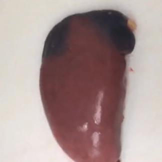
Spleen dissection.
-
Case 9 – history
Spleen dissection.
-
Case 8 Part 6
Inferior vena cava. TIPS device (shunt).
-
Case 8 Part 5
Aorta assessment. Dual superior mesenteric artery ostia.
-
Case 8 Part 4
Pleural fluid. Pericardial fluid. Splenomegaly. Bowel infarct.
-
Case 8 Part 3
Bowel infarct.
-
Case 8 Part 2
External exam. Peritoneal dialysis catheter. Central venous access sites. Long bone donation.
-
Case 8 Part 1
TIPS fluoroscopy. Postoperative CT scan.
-
Case 8 – History
TIPS procedure.
-
Case 7 Part 6
Heart and lung block.
-
Case 7 Part 5
Inferior vena cava filter.
-
Case 7 Part 4
Ureter. Inferior vena cava.
-
Case 7 Part 3
Initial dissection.
-
Case 7 Part 2
Y-shaped incision.
-
Case 7 Part 1
Rigor mortis.
-
Case 7 – History
Inferior vena cava filter.
-
Case 6 Part 8
Heart anatomy.
-
Case 6 Part 7
Abscess.
-
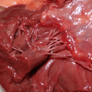
Case 6 Part 6
Spinal cord. Cauda equina.
-
Case 6 Part 5
Right flank assessment.
-
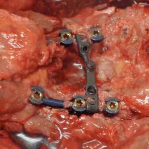
Case 6 Part 4
Spinal cage.
-
Case 6 Part 3
Bone saw.
-
Case 6 Part 2
Spine exposure.
-
Case 6 Part 1
External exam. Multiple surgical scars.
-
Case 6 – History
Spinal surgery.
-
Case 5 Part 3
Cerebral anatomy.
-
Case 5 Part 2
Cerebellar anatomy.
-
Case 5 Part 1
Base of skull anatomy.
-
Case 5 – History
Dementia
-
Case 4 Part 3
Tumor attached to pericardium. Pleural effusion.
-
Case 4 Part 2
Transdiaphragmatic tumor. Pleural effusion.
-
Case 4 Part 1
External exam.
-
Case 4 – History
Transdiaphragmatic tumor.
-
Case 3 Part 11
Left dominant coronary artery system.
-
Case 3 Part 10
Assessment of coronary arteries.
-
Case 3 Part 9
Airway. Aorta. Esophagus.
-
Case 3 Part 8
Lung sampling for microscopy.
-
Case 3 Part 7
Lung weights. Pulmonary congestion and edema.
-
Case 3 Part 6
Pulmonary apical blebs (emphysema).
-
Case 3 Part 5
Organ block. Chest cavity.
-
Case 3 Part 4
Continued chest assessment.
-
Case 3 Part 3
Initial chest assessment.
-
Case 3 Part 2
Y-shaped incision.
-
Case 3 Part 1
External exam.
-
Case 3 – History
Recent chest pain.
-
Case 2 Part 4
Laryngeal stenosis. Traumatic intubation.
-
Case 2 Part 3
Severe 2-vessel coronary artery disease.
-
Case 2 Part 2
Aspiration.
-
Case 2 Part 1
Gastrostomy tube. Jejunostomy tube.
-
Case 2 – History
Diabetes, stroke, feeding tube.
-
Case 1 Part 13
Microscopy — pulmonary edema.
-
Case 1 Part 12
Lining of aorta. Left anterior descending coronary artery (plaque).
-
Case 1 Part 11
Airway, heart (external), esophagus — basic anatomy.
-
Case 1 Part 10
Lung sectioning.
-
Case 1 Part 9
Heart and lung block — basic anatomy.
-
Case 1 Part 8
Removal of bladder. Status post hysterectomy. Basic survey of abdomen. Scoliosis.
-
Case 1 Part 7
Removal of intestine.
-
Case 1 Part 6
Neck anatomy — basic review. Removal of chest organs.
-
Case 1 Part 5
Assessment for pulmonary embolism.
-
Case 1 Part 4
Removal of chest plate.
-
Case 1 Part 3
Abdominal survey — basic anatomy. Adhesions.
-
Case 1 Part 2
Y-shaped incision. Livor mortis within subcutaneous tissue.
-
Case 1 Part 1
External exam. Anterior livor mortis. EKG leads.
-
Case 1 – History
Recent pneumonia.
-
Histology Portal
-

StayTuned